cortical thickness measurement|normal cortical thickness of kidney : distributor The thickness of the motor cortex is often underestimated in MRI thickness measurement , probably because of the high degree of intracortical myelination that affects the gray–white contrast, causing the white matter surface to be too close to the gray surface, such that cortical thickness is underestimated [10,14]. The calcarine sulcus is . Meu marido adora apertar e mamar neles 😈🔥🔥. 35 upvotes · 4 comments. 142K subscribers in the brgonewild community. Paraíso das musas tupiniquins!
{plog:ftitle_list}
WEB3D Warehouse is a website of searchable, pre-made 3D models that works seamlessly with SketchUp. We use web browser cookies to create content and ads that are relevant to you. By continuing to use this site, you are consenting to our cookie policy. You can also manage cookie preferences.
why is cortical thickness important
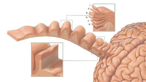
The cortical thickness is then calculated at every volumetric point within the cortex and is based on the length of the trajectory from one boundary to another. Here we investigate a method for . Cortical thickness in neuroimaging is a common measure used to describe the distance between the innermost and outermost edges of the cerebral cortical gray matter (see Fig. 1).This can then be used to describe an average thickness for the entire brain (i.e., global), or it can be localized (i.e., local) to different regions of interest (e.g., dorsolateral prefrontal .The cortical thickness has been used as a biomarker to assess different cerebral conditions and to detect alterations in the cortical mantle. In this work, we compare methods from the FreeSurfer software, the Computational Anatomy Toolbox (CAT12), a Laplacian approach and a new method here proposed, based on the Euclidean Distance Transform (EDT), and its corresponding .
These differences in clustering results are attributable at least in part to the many methodological differences between the two studies, such as multi-scanner data in the Karama et al. (2009) study, different methods of extraction of the cortical surface and measurement of cortical thickness, and different techniques to control p-values over . The thickness of the motor cortex is often underestimated in MRI thickness measurement , probably because of the high degree of intracortical myelination that affects the gray–white contrast, causing the white matter surface to be too close to the gray surface, such that cortical thickness is underestimated [10,14]. The calcarine sulcus is .
selected tear film tests in healthy cats
Background Cortical thickness measures the width of gray matter of the human cortex. It can be calculated from T1-weighted magnetic resonance images (MRI). In group studies, this measure has been shown to correlate with the diagnosis/prognosis of a number of neurologic and psychiatric conditions, but has not been widely adapted for clinical routine. One of the .Acosta O, Bourgeat P, Zuluaga MA et al (2009) Automated voxel-based 3D cortical thickness measurement in a combined Lagrangian-Eulerian PDE approach using partial volume maps. Med Image Anal 13:730–743 Lerch JP, Carroll JB, Dorr A et al (2008) Cortical thickness measured from MRI in the YAC128 mouse model of Huntington’s disease.
Laplace methods [Haidar and Soul,2006; Jones et al.,2000; Yezzi and Prince,2003] solve Laplace's equation in the GM region with the boundary condition of constant (but different) potentials on each of the two surfaces.The cortical thickness is then defined at each point as the length of the integral curve of the gradient field passing through that point, as . Cortical thickness measurements are then estimated using the DiReCT algorithm [Das et al., 2009]. Briefly, the DiReCT method estimates the GM/WM interface and the GM/CSF interface and computes a diffeomorphic mapping between the two interfaces, from which thickness is derived. We note that this is performed in native space and no correction for .Cortical thickness should be estimated from the base of the pyramid and is generally 7–10 mm. . Chronic renal disease caused by glomerulonephritis with increased echogenicity and reduced cortical thickness. Measurement of kidney length on the US image is illustrated by ‘+’ and a dashed line. Figure 23. Open in a new tab.
We would like to show you a description here but the site won’t allow us.Measuring Thickness in MRI with AFNI [1] Fischl B, Dale AM, 2000, PNAS, 97,20,11050-11055 [2] Das SR,et al. 2009, NeuroImage, 45,867-879 . Thickness, especially cortical thickness, is a widely used metric in MRI, yet there is little in a gold standard to establish its veracity. Common methods (e.g. FreeSurfer) rely on complex procedures that . Cortical thickness is an important biomarker for image-based studies of the brain. A diffeomorphic registration based cortical thickness (DiReCT) measure is introduced where a continuous one-to-one correspondence between the gray matter–white matter interface and the estimated gray matter–cerebrospinal fluid interface is given by a diffeomorphic mapping in the . Measurement of cortical thickness index (CTI) and canal flare index (CFI) on an anteroposterior radiograph (using Osirix software). Do: outer diameter (the shaft's outer diameter at 10 cm below the lesser trochanter), Di: inner diameter (the shaft's inner diameter at 10 cm below the lesser trochanter, measured at the same level as Do), CW .
Sonographic cortical bone thickness measurement. The ultrasonographic CoT values of each participant were measured using the same ultrasound device (Toshiba Aplio 500; Japan) and transducer. The measurements were taken by the same radiologist from the radius, tibia, and anterior cortical areas of the second metatarsal head of the non-dominant .
After cortical thickness measurement, we found that there is a significant decrease in the cortical thickness in the RRMS group compared to the control group in the mean cortical thickness of the left and right hemispheres, and found focal thickness reduction in precentral, paracentral, postcentral and posterior cingulate cortices in both . In this paper, we investigate scanner effects in two large multi-site studies on cortical thickness measurements across a total of 11 scanners. We propose a set of tools for visualizing and identifying scanner effects that are generalizable to other modalities. We then propose to use ComBat, a technique adopted from the genomics literature and .For example, used FreeSurfer cortical thickness measurements across image acquisition sessions to demonstrate improved reproducibility with the longitudinal stream over the cross-sectional stream. In comparisons for ANTs, FreeSurfer, and the proposed method were made using the range of measurements and their correspondence to values published . The cerebral cortex is a highly folded outer layer of grey matter tissue that plays a key role in cognitive functions. In part, alterations of the cortex during development and disease can be captured by measuring the cortical thickness across the whole brain. Available software tools differ with regard to labor intensity and computational demands. In this study, we .
Cortical Thickness: Measurement of the thickness of the cerebral cortex, providing insight into neurological and psychiatric conditions. Importance in Medicine: Indicates brain development, aging, and health, and aids in diagnosing conditions like Alzheimer's and schizophrenia. In addition, a 3D color map uses differences in color to denote the thickness of the cortical bone in the segmented output. Through this algorithm, the cortical and marrow bones in the TMJ condyle head were segmented with high accuracy, and the measured cortical thickness was visualized as a 3D-rendered color map. Introduction. Measuring the thickness of the human cerebral cortex is of great interest in studies of both normal and abnormal neuroanatomy. The average thickness over the whole brain is around 2.5 to 3 mm and within individual brains varies from about 2 mm at its thinnest in the calcarine cortex up to 4 mm and over in the thicker regions of the precentral .
cortical thickness with a color display based on the segmentation results. In total, 12,800 CBCT images from 25 normal subjects, manually labeled by an oral radiologist, served as the gold‑standard. the time of examination. Renal length measurements were performed in the sagittal view, and the maximum length of each kidney was measured. Measurements of parenchymal thickness and medullary pyramid thickness were performed on the same image on which length mea-surements were made. Parenchymal thickness and medullary pyramid .
what does cortical thickness indicate
normal cortical thickness of kidney
Objective: The purpose of our study was to determine whether there is a relationship between renal cortical thickness or length measured on ultrasound and the degree of renal impairment in chronic kidney disease (CKD). Materials and methods: From October to December 2007, 25 patients (13 men and 12 women, mean age 73 years) were identified who had CKD but were .
Recently, some cortical thickness measurement methods have been developed based on these chosen methods aiming to remove particle volume effects and improve thickness measurement in gyri and sulci (Acosta et al., 2009; Bourgeat et al., 2008). In this study, we performed two analyses, one with standard defaults and one with corrections of the . Similarly, Tingart et al. concluded measurement of cortical thickness of the proximal humerus is reproducible and a thickness <4 mm is highly indicative of low BMD. In the current study, we select to study postero-medial thickness of the tibia as it is a weight bearing bone that should better correlate prediction of osteoporosis. The distribution of cortical bone in the proximal femur is believed to be a critical component in determining fracture resistance. Current CT technology is limited in its ability to measure cortical thickness, especially in the sub-millimetre range which lies within the point spread function of today’s clinical scanners.
self acl tear test

Resultado da 1.3M. Marque seus amigos que queriam ter um chefe desse 😈. Japinha (@nordestinajapa_) no TikTok |1.2M curtidas.410.7K seguidores.@japanordestina Faço uns vídeos maneiros que você vai gostar de assistir ️🔥.Assista ao último vídeo de Japinha (@nordestinajapa_).
cortical thickness measurement|normal cortical thickness of kidney