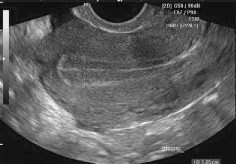
how to measure endometrial thickness on ultrasound|3 line sign endometrium ultrasound : advice How to measure endometrial thickness (ET) 1. When intracavitary fluid is present, measure thickness of both single layers and add together to give ET 2. When intracavitary pathology is . Resultado da BRJOGOS is the best online gaming platform in Brazil, where you can play hundreds of games and win amazing prizes. Join now with the affiliate .
{plog:ftitle_list}
WEB8 de fev. de 2024 · Masha is a little girl with the face of an angel who lives hell in the life of her best friend, a huge and sweet retired bear from the circus. He tries to keep Masha out of trouble but often.

Ultrasound measurements of endometrial thickness were originally carried out by measuring the distance from anterior stratum basalis to the posterior stratum basalis, and dividing by 2 to .
Review the ideal technique to measure endometrial thickness while performing pelvic ultrasound. Review how ultrasound can be used to characterize adnexal masses in those who are postmenopausal. Access free . Based on this analysis, and in comparison with the threshold that is widely accepted in women with bleeding, an endometrial thickness measurement of ≥ 11 mm .
The endometrial thickness is the maximum measurement in the sagittal plane and includes both endometrial layers (double endometrial thickness). It is critical to ensure that the uterus is in a .
How to measure endometrial thickness (ET) 1. When intracavitary fluid is present, measure thickness of both single layers and add together to give ET 2. When intracavitary pathology is .Normal range of endometrial thickness. The designation of normal limits of endometrial thickness rests on determining at which thickness the risk of endometrial carcinoma is .Moreover, endometrial thickness can vary with the menstrual cycle and with the use of hormone replacement therapy or selective estrogen receptor modulators. In this review, the use of .To determine the accuracy of endometrial thickness measurement by pelvic ultrasonography in the diagnosis of endometrial carcinoma and disease (hyperplasia and/or carcinoma) in .
How to measure endometrial thickness (ET) 1. When intracavitary fluid is present, measure thickness of both single layers and add together to give ET 2. When intracavitary pathology is present measure total ET including the lesion (unless it’s a well defined myoma that can be measured separately) Leone et al. UOG, 2010, 35: 103–112 The uterine lining is called the endometrium. During an imaging test, it’ll show up as a dark line. This is the “endometrial stripe.” Here’s how this tissue can change with age, symptoms .
secretory phase (day 16 to 28): thickened hyperechoic endometrium measuring up to 16 mm; Postmenopausal women: regular, thin hyperechoic line measuring up to 5 mm, representing the remaining basal layer of endometrium; Please see the separate article on endometrial thickness for a detailed discussion of measurements and pathological .
trilaminar endometrium ultrasound images
normal endometrial thickness on ultrasound
custom gain express ph & soil moisture meter
Endometrial thickness is measured in millimeters using an ultrasound or magnetic resonance imaging (MRI). "Normal" endometrial thickness measurements are as follows during the various phases: . Surgery may also be considered if the tissue impacting endometrial thickness is structural, such as a fibroid or polyp. In these cases, surgery may .Although every ultrasound examination should still start with a correct measurement of the endometrium, especially in those with a thickened endometrium the above mentioned ultrasound applications should be used to reach a more precise diagnosis (e.g. an endometrial polyp with the typical presence of a pedicle artery on colour Doppler; an . In addition to measuring the thickness, transvaginal ultrasound also makes it possible to analyze another important parameter for implantation purposes, namely the appearance of the endometrium . Depending on the phase of the menstrual cycle, in fact, the inner lining of the uterus changes and takes on the following ultrasound characteristics:
Introduction. Postmenopausal vaginal bleeding is a common complaint and is associated with a 1–10% risk of endometrial cancer, depending on age and risk factors 1, 2.Because the risk of cancer is relatively high, the clinical standard of care requires diagnostic evaluation to exclude malignancy 2, 3.Until the 1980s, fractional dilation and curettage was the . During transvaginal ultrasound evaluation, endometrial thickness, echo pattern, and endometrial perfusion are evaluated. . However, an endometrial thickness measurement of 7 mm or greater may warrant further evaluation to rule out any potential underlying health issues, particularly if there are other concerning symptoms or factors present . Ultrasound is the first-line imaging test to evaluate the endometrium. The normal endometrium is composed of 2 layers and the combined thickness of the 2 layers depends on where a woman is in her menstrual cycle (Figure 1, Figure 2, Figure 3) [1].The best way to measure the endometrial thickness is on a midsagittal transvaginal image.
Purpose: Endometrial thickness is one of the most important indicators in endometrial disease screening and diagnosis. Herein, we propose a method for automated measurement of endometrial thickness from transvaginal ultrasound images. Methods: Accurate automated measurement of endometrial thickness relies on endometrium . An ultrasound scan. An ultrasound scan is usually arranged if a doctor suspects endometrial hyperplasia symptoms. This can check for other causes of bleeding, such as polyps (benign fleshy lumps) in the womb (uterus), or cysts on the ovaries. The scan can also measure the thickness of the womb lining.
Gulfcoast Ultrasound Instructor Brian Schenker, MBA, RDMS, RVT explains how to measure the endometrium during a routine pelvic exam using ultrasound.Upcoming.
Endometrial thickness varies according to a woman's age and menstrual cycle. A healthy endometrium is essential for a healthy pregnancy. An endometrial thickness of less than 14 mm is typically considered normal at any stage of the menstrual cycle. During menstruation, the endometrial thickness of pre-menopausal women ranges between two and .Zoom the image to assess and measure the endometrial thickness. Rotate into transverse and angle slightly cranially to be perpendicular to the uterus. Whilst in transverse and slightly right of midline, angle left laterally to identify the left ovary using the full bladder as an acoustic window.Ultrasound measurement. Measurement of endometrial thickness and pattern was performed 11–12 hours before the hCG injection by transvaginal 8 MHz ultrasonography with Doppler Ultrasound (Mindray DC-6 Expert, Shenzhen, China) after patients had rested for at least 15 minutes and completely emptied their bladders. Endometrial thickness was .
A transvaginal ultrasound exam may be done to measure the thickness of the endometrium. For this test, a small device is placed in your vagina. Sound waves from the device are converted into images of the pelvic organs. If the endometrium is thick, it may mean that endometrial hyperplasia is present. Endometrial assessment includes measurement of endometrial thickness, identifying the endometrial morphological pattern, and grading the endometrial vascularity. To assess endometrial vascularity, the color flow . The measurement of endometrial thickness, however, is associated with a 1% false-negative rate and there are reports in the literature of advanced endometrial cancer in patients with an endometrial thickness of < 5 mm 3, 4. Endometrial hyperplasia: It is diagnosed by measuring combined endometrial thickness. There may be focal or diffuse endometrial thickening with or without internal cystic spaces. . On ultrasound, an interrupted endometrium line is noted in the sagittal plane with punctate echogenic foci within the endometrium. Echogenic fibrotic septae with .
Quantitative assessment of endometrial thickness: (Figures 4-7) The endometrial thickness is the maximum measurement in the sagittal plane and includes both endometrial layers (double endometrial thickness). It is critical to ensure that the uterus is in a midsagittal plane, the whole endometrial stripe is seen from the fundus to the endocervix .
Measurement of the endometrium can be used to help assess for pathologies such as hyperplasia or neoplasm that may expedite referral to gynecology for further evaluation or biopsy. The table below outlines the normal values for endometrial thickness which generally decreases after menopause (Nalaboff et al., 2001; Ozcan & DeCherney, 2019). Problems with a rigid application of cut-offs to ultrasound measurements of endometrial thickness were also recognized by Leung et al. 10, who concluded that a combination of clinical and ultrasound findings should be used to establish the diagnosis of incomplete miscarriage and plan further management.
Retained products of conception can be suspected on ultrasound if the endometrial thickness is >10 mm following dilatation and curettage or spontaneous abortion (80% sensitive) 13. Furthermore, if the endometrial thickness is less than 10 mm, there is a relatively high negative predictive value (63-80%) for RPOC 13 .The designation of normal limits of endometrial thickness rests on determining at which thickness the risk of endometrial carcinoma is significantly increased. Whilst quantitative assessment is important, endometrial morphology and the presence of risk factors for endometrial malignancy should also be taken into account when deciding whether or .
To be included in the review, studies had to measure endometrial thickness using ultrasound imaging. One or both layers of the endometrium were measured; the cut-offs for an abnormal test ranged from less than or equal to 2 mm to less than or equal to 10 mm for single-layer measurement, and from 3 to 15 mm for the measurement of both layers.
How To Measure Uterus On Ultrasound | Uterine Length, Width, AP Thickness Measurements TA/TVS USGUterus Measurements:Length: 7.5cm (approximately)Width: 5cm .
custom gain express ph & soil moisture meter 295mm
endometrium thickness chart stages ultrasound
Resultado da 2 de jan. de 2024 · Pensando nisso, o NG+ prepara todo mês uma lista com todos os códigos ativos e válidos para .
how to measure endometrial thickness on ultrasound|3 line sign endometrium ultrasound