long bone compression test foot|metatarsal fracture testing pdf : discount store
WEBOur free horse racing tips feature everything from National Hunt racing to Flat racing, across a range of distances at a variety of tracks. These tips are provided to help you narrow .
{plog:ftitle_list}
webA lot of you have been spamming this sub with non sense. Going forward, it will be an immediate ban from this sub. We have rules for this sub for a reason so I highly suggest .
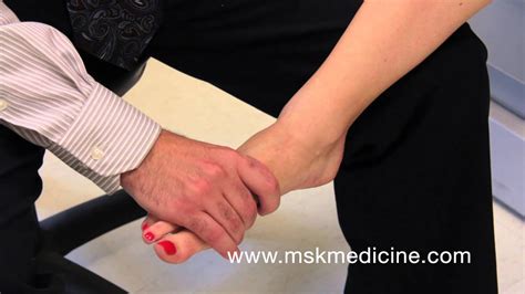
The Long Bone Compression Test is an orthopedic special test utilized in evaluation of suspected foot injury. www.whitworth.edu/msat.Purpose: To assess for a fracture in the lower extremity. Test Position: Supine. Performing the Test: The patient should not be wearing shoes. The examiner then strikes the heel of the patient. A positive test is reproduction of the .The subject sits with the affected leg extended and the foot off the end of the examining table. The examiner stands at the end of the table near the subject's foot. Foot drop is sometimes caused by a mass pushing on a nerve. This can be an overgrowth of bone in the spinal canal or a tumor or cyst pressing on the nerve in the knee or .
metatarsal squeeze test forefoot
metatarsal loading tests pdf
In this video I will be demonstrating how to perform the Long bone compression for the foot.
Foot drop (also called drop foot) happens when you can’t raise the front part of your foot due to weakness or paralysis of the muscles that lift it. It’s a symptom of several possible underlying .
For example, if the stress fracture is in your leg or foot, you can prop your leg up with pillows or cushions while you’re lying down. Compression: Compression helps reduce blood flow to your injured bone and reduces .
Use pneumatic compression device (e.g., a stirrup leg brace, compression walking boots) or other biomechanical stress-relieving measures (e.g., crutches) for lower-extremity stress fractures 20.Importance of Test: Our bones are covered by a layer of tissue known as the periosteum. It is highly innervated and very sensitive to injury. When a bone is fractured, the periosteum is more easily stimulated and thus pain is .Test Position: Supine. Performing the Test: . When a bone is fractured, the periosteum is more easily stimulated and thus pain is experienced. By striking the heel of the foot, a large vibratory/compression force is sent through the limb .
long bone compression test pain or increased glide when compared to same joint on opposite extremity pt: supine w/ knee flexed and heel stabilized by table examiner: standing in front of pt's feet, one hand grasping distal tarsal row (cuneiforms) and other hand grasping the mt being glided procedure: glide mt dorsally on the tarsal then .
Guidelines for splinting long-bone injuries include: A. assessing the radial pulse for a lower extremity injury. B. splinting the injured extremity to the uninjured extremity. C. immobilizing the hand or foot in the position of function. D. having the .Long bone compression test. Tarsal Tunnel Syndrome: Tests. Tinel's Sign. Intermetatarsal Neuroma: Tests . in supine position - Examiner stabilizes the talus superiorly while gripping the calcaneous at the plantar aspect of foot - The examiner applies a medial glide of calcaneous on the fixed talus. Subtalar Joint Play: Postive test. A broken foot, or foot fracture, can affect any of the 26 bones in each foot. Depending on your injury, recovery time for a fractured foot can vary from six weeks to six months. . An X-ray is the most common diagnostic test used to diagnose a foot fracture. The Ottawa Ankle and Foot Rules are used as a screening measure to determine if an X .760 | september 2012 | volume 42 | number 9 | journal of orthopaedic & sports physical therapy [research report]S tress fractures are a bone-related overuse injury commonly seen in athletes and military personnel.29,35 This condition was first reported in the literature in 1855 by a Prussian military phys-
About Press Copyright Contact us Creators Advertise Developers Terms Privacy Policy & Safety How YouTube works Test new features NFL Sunday Ticket Press Copyright .The bones of the feet are commonly divided into three parts: The hindfoot; The midfoot; The forefoot; Seven bones — called tarsals — make up the hindfoot and midfoot. The calcaneus (heel bone) is the largest of the tarsal bones in the foot. It lies at the back of the foot (hindfoot) below the three bones that make up the ankle joint. Compression fractures can happen to any part of your spine, but they usually occur in the thoracic spine (middle section). Osteoporosis is a common cause of compression fractures, in addition to trauma (like after an accident) or tumors that weaken the bone.. A healthcare provider may treat these fractures with medications, a back brace or surgery, .
metatarsal fracture testing pdf
Stress injuries represent a spectrum of injuries ranging from periostitis, caused by inflammation of the periosteum, to a complete stress fracture that includes a full cortical break. They are relatively common overuse injuries in athletes that are caused by repetitive submaximal loading on a bone over time. Stress injuries are often seen in running and jumping athletes .
Bone healing is a natural process. Our bone is constantly being replaced with new bone, and after a bone injury occurs, the body has a tremendous capability to heal the damage to the bone. People who sustain broken bones typically will heal these fractures with appropriate treatment that may include casts, realignment, and surgery. Medial malleolus: The bony bump on the inner side of the ankle.; Talus: An ankle bone located at the top and back of your foot.This is where the two lower leg bones (tibia and fibula) connect to the foot. Tarsal navicular: A small bone in the midfoot that has an important role in maintaining the arch of the foot.; Proximal fifth metatarsal: One of the five long, thin .
Ankle Clearing Test: . with the knee bent, ankle is placed in full plantarflexion. The examiner applies compression and scours the joint. Then, with the knee bent, ankle is placed in full dorsiflexion. The examiner applies compression and scours the joint. . or the navicular bone (for foot injuries) Note: tests should only be performed by a .
A stress fracture in the foot is an overuse injury. It's common in athletes and people who try to do too much activity too quickly. Learn how to recognize signs of a stress fracture.Long Bone Compression Test. How: Patient is lying with knee extended, apply a longitudinal force along the shaft of the bone. . How: Tap along the path of the posterior tibial nerve Positive Test: N&T into the foot and toes Indicates: Posterior tibial nerve dysfunction OR . Self-adherent compression bandages, such as Coban or Dynarex, are bandages that behave like tape but do not stick to the skin. They can be torn to specific lengths and come in widths ranging from 1/2 to 4 inches. Self . A bone stimulator is a device that generates an electric current meant to encourage bone growth. It uses ultrasonic or pulsed electromagnetic waves. To be effective, bone stimulator treatment must .
Foot radiography is required if there is pain in the midfoot zone and any of the following: bone tenderness at point C (base of the fifth metatarsal) or D (navicular), or inability to bear weight .The talus is one of the bones in the heel of the foot. It is an uncommon bone to be affected by stress fracture. When it does occur, however, it can cause pain in the heel or ankle. Stress Fracture of the Sesamoids. The sesamoids are two small bones located in the ball of the foot, beneath the joint of the big toe.Triple Compression Stress Test: Place patient’s foot in full plantarflexion and inversion with one hand, while simultaneously applying digital pressure over the tarsal tunnel just posterior to the media malleolus for 30 seconds. The test is considered positive if patient reports new paresthesia, pain, or numbness in the posterior tibial nerve .Stress Test: Long Bone Compression Test. Patient Position: supine with affected leg extended. ankle and foot just off table Examiner Position: next to patients leg and notes where the pain is . Eval. Procedure: knee of invovled leg is fully extended, the examiner passively dorsiflexes the subjects foot + Test: pain in calf when the foot is .
X-rays can help rule out a fracture or other bone injury as the source of the problem. Magnetic resonance imaging (MRI) also may be used to help diagnose the extent of the injury. . X-ray; Treatment. For immediate self-care of a sprain, try the R.I.C.E. approach — rest, ice, compression, elevation: Rest. Avoid activities that cause pain . Foot fractures are so common that one out of every ten cases of broken bone injuries occur in the foot. Discover how compression socks help in healing foot injuries. FREE SHIPPING WITHIN THE USA! . The forefoot is the long front part of the foot, consisting of nineteen bones. . The talus is a bone in the ankle that connects the lower leg to .Anatomy-Foot Bones. 16 terms. tessajmcgowan. Preview. Reading - Ped = foot. 10 terms. ros_salguero. Preview. Dermatomes. 17 terms. jisooz. Preview. Foot, Ankle, Lower Leg Special Tests. . Long bone compression test. Place one hand around proximal foot/ankle to stabilize Push proximally on metatarsal head. Tap test. Tap end of toe. Foot drop is a general term that describes a difficulty in lifting the front part of the foot. It's often caused by compression of a nerve. . This test uses radio waves and a strong magnetic field to create detailed images of bones and soft tissues. . nerve surgery might be helpful. If foot drop is long-standing, your doctor might suggest .

A back bone consists of a drum-shaped part (body) in the front, a hole for the spinal cord, and several projections of bone (called processes) in the back. Disks of cartilage between each back bone help cushion and protect the bones. In compression fractures, the body of a back bone collapses, usually because of too much pressure.
WEB360p. 1. 3 min Admirador-De-Gordinhas-Bbw - 751.8k Views -. Show more related videos. XVIDEOS Mulheres cheinhas e deliciosas free.
long bone compression test foot|metatarsal fracture testing pdf