testing for a tear in the retina|how to treat retinal tear : solutions Retinal detachment may cause you to lose vision. Depending on your amount of vision loss, your lifestyle might change a lot. You may find the following ideas useful as you . See more webThe latest tweets from @lays_rezende
{plog:ftitle_list}
Resultado da ASSINE MEUS VÍDEOS DO SITE👇 acompanhanteyumi.com/seja-premium. TWITTER OFICIAL 👇 twitter.com/yumi_villar. Meu INSTAGRAM OFICIAL 👇 .
signs of a torn retina
Diagnosis involves the steps that your healthcare professional takes to find out if retinal detachment is the cause of your symptoms. Your healthcare team may use the following tests and instruments to diagnose retinal detachment: 1. Retinal exam.Your healthcare professional may use an instrument with a bright light . See moreSurgery is almost always the type of treatment used to repair a retinal tear, hole or detachment. Various techniques are available. Ask your ophthalmologist about the . See more
Retinal detachment may cause you to lose vision. Depending on your amount of vision loss, your lifestyle might change a lot. You may find the following ideas useful as you . See more
Cardamom Powder moisture meter
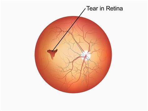
A retinal tear or break happens when the gel-like vitreous in your eye pulls on your retina and causes a split. Your retina is a thin layer of tissue that’s sensitive to light found at the back of your eye. A torn retina is a serious problem that makes your vision blurry. It is when the retina has a tear or hole, like a rip in cloth. A torn retina often leads to a more serious condition . However, prompt treatment is needed. A retinal tear can quickly progress to a retinal detachment, which can cause permanent vision loss. This article discusses retinal tears. It examines the symptoms, causes, and . The following tests may be done to find the location and extent of the disease: Amsler grid test. An eye professional may use an Amsler grid to test the clarity of your central vision. You'll be asked if the lines of the grid seem .
A retina specialist will check for retinal tears by placing drops in your eyes to dilate the pupil. They’ll look through a special lens to assess any changes inside the eye. Eye surgery is the most common treatment for a torn . A retinal tear is a rip in the layer of light-detecting cells at the back of your eye called the retina. It’s a medical emergency that can lead to permanent vision loss if not treated.
A thorough and timely examination by a retina specialist using scleral depression (applying slight pressure to the eye) and/or a 3-mirror lens is the most important step in diagnosing a . An eye doctor can check for a retinal detachment or retinal tears with a dilated eye exam. During this test, they give you eye drops to widen your pupil and examine the back of your eye.
Retinal tears can develop when the vitreous gel separates from the retina as part of aging or in patients with abnormal thinning in the peripheral retina (known as lattice degeneration) or . Li, KX, et al. “ Contemporary Management of Complex and Non-Complex Rhegmatogenous Retinal Detachment Due to Giant Retinal Tears.”Clinical Ophthalmology, 2021. Ghosh, YK, et al. “ Surgical treatment .You'll be referred to hospital for surgery if tests show your retina may be detached or has started to come away (retinal tear). . sealing the tear in your retina with laser or freezing treatment (cryotherapy) It's usually done with local anaesthetic, so you're awake but your eye is numbed. had a serious eye injury; had a retinal tear or detachment in your other eye; have family members who had retinal detachment; have weak areas in your retina (seen by an eye doctor during an exam) Early Signs of a Detached Retina. A detached retina has to be examined by an ophthalmologist right away. Otherwise, you could lose vision in that eye.
The Schirmer's test is used to find out if a person is producing enough tears. Without moisture, the eyes can become dry, increasing the risk of eye health problems. A piece of paper is placed in .This device lowers eye pressure by allowing excess fluid to escape the eye. A vitrectomy removes the vitreous that might otherwise plug the drainage tube. In each example, a vitrectomy improves the outcome of the procedure and reduces the likelihood of retinal tear, retinal detachment, macular edema (swelling), and other complications. 4.
Schirmer's test determines whether the eye produces enough tears to keep it moist. This test is used when a person experiences very dry eyes or excessive watering of the eyes. It can cause damage to the cornea. [1] A negative (more than 10 mm of moisture on the filter paper in 5 minutes) test result is normal.
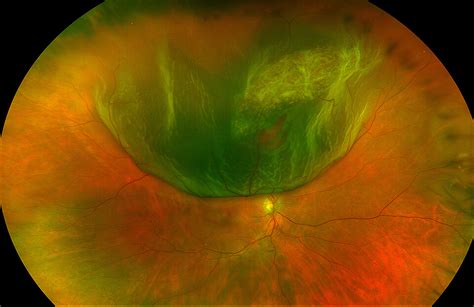
A small tear in your retina lets the gel-like fluid called vitreous humor travel through the tear and collect behind your retina. The fluid pushes the retina away, detaching it from the back of your eye. . Your provider may recommend other tests after the dilated eye exam. These tests are noninvasive. They won’t hurt. They help your .
A retinal tear is a rip in the layer of light-detecting cells at the back of your eye called the retina. It’s a medical emergency that can lead to permanent vision loss if not treated quickly. The doctor may press on your eyelids to check for retinal tears, which may be uncomfortable for some people. . Both of these tests are painless and can help your eye doctor see the exact position of your retina. What’s the treatment for retinal detachment? Depending on how much of your retina is detached and what type of retinal detachment .Most commonly utilized diagnostic tests for dry eye and some of the newer tests and their techniques were summarized below. Tear Film Stability. Fluorescein BUT: The tear break-up time test is easily and quickly performed via fluorescein dye instillation. After the fluorescein is instilled, the patient is asked to stare without blinking. Osmolarity Testing Osmolarity instruments are used to test all kinds of solutions—blood, serum, plasma, urine, bilirubin, milk, cell culture media, and many others. In the world of research, tears are almost among the least of these. Still, three different diagnostic technologies have been used for tear film osmolarity testing.

Retinal tears may occur as people age when their vitreous shrinks and pulls the retina from the back of the eye. If a tear occurs, a person may notice a sudden increase in floaters, dark spots in .The Amsler Grid test can be an important indicator of diseases in the retina. Test your eyes daily to detect changes as early as possible. If you normally wear reading glasses, please wear them while performing this test. Proper lighting also is essential. The Amsler Grid should be held at a normal reading distance and should be tested from the . Retinal tears occur when the vitreous gel inside the eye pulls away from the retina, causing a tear or hole in the delicate tissue. This can lead to a variety of symptoms, including floaters, flashes of light, and a sudden decrease in vision. . Before laser treatment, patients may need to undergo a thorough eye examination and imaging tests .
Diagnostic testing. A thorough and timely examination by a retina specialist using scleral depression (applying slight pressure to the eye) and/or a 3-mirror lens is the most important step in diagnosing a retinal tear. In cases where .Retinal tears can develop when the vitreous gel separates from the retina as part of aging or in patients with abnormal thinning in the peripheral retina (known as lattice degeneration) or occasionally from trauma. . Diagnostic testing. Your retina specialist will perform a detailed eye exam, including a careful examination of the peripheral .
This type of retinal detachment is the most common. A rhegmatogenous detachment is caused by a hole or tear in the retina that lets fluid pass through and collect underneath the retina. This fluid builds up and causes the retina to pull away from underlying tissues. The areas where the retina detaches lose their blood supply and stop working. Retinal tear. A retinal tear occurs when the clear, gel-like substance in the center of your eye, called vitreous, shrinks and tugs on the thin layer of tissue lining the back of your eye, called the retina. This can cause a tear in the retinal tissue. It's often accompanied by the sudden onset of symptoms such as floaters and flashing lights.
A retinal tear is diagnosed through a comprehensive eye exam, including a dilated eye exam and imaging tests such as optical coherence tomography (OCT) and fluorescein angiography. Retinal tear surgery involves sealing the tear with laser or cryotherapy to prevent further damage to the retina and preserve vision. TearLab Osmolarity Testing The TearLab test measures how concentrated the electrolytes in the tears are, with higher levels signifying that the aqueous component of the tears is low. To administer the test, the physician or technician takes a 50-µl sample of a patient’s tears from each eye using a special pen and sample card.
Diagnostic testing. A thorough and timely examination by a retina specialist using scleral depression (applying slight pressure to the eye) and/or a 3-mirror lens is the most important step in diagnosing a retinal tear. In cases where there is a limited view of the retina due to overlying hemorrhage, ophthalmic ultrasound may be required to aid .
Retinal Tears and Holes. Retinal breaks are defined as any full thickness tears or holes in the retina. They are key risk factors for retinal detachments. Retinal tears are usually caused by traction on the retina from a PVD. Retinal holes are commonly caused by vitreous traction or lattice degeneration creating an atrophic hole. SymptomsHowever, a sudden increase of floaters, which might look like a shower or a curtain of floaters, signifies a retinal tear or detachment. Other warning signs of a retinal tear include flashes of light, shadows or veils over your vision, sudden blurry vision, or decreased peripheral vision. If you notice any of these symptoms, call us right away .Use the Amsler grid in the same way each time you test your vision. If you normally wear reading glasses, please wear them while performing this test. Good lighting is essential. 1) Hold the Amsler grid at a normal reading distance. 2) Test each eye separately. Close one eye or cover it with your hand 3) Look at the dot in the center of the grid. This is another test that can help a doctor check for adequate tear production. For this test, numbing eye drops will be placed in your eye, and a small piece of paper will be placed on the edge .

Turmeric Powder moisture meter
A quick test called optical coherence tomography (OCT) is the best way to diagnose a hole in the retina. . Some people use the words “retinal hole” and “retinal tear” to mean the same .
Diagnosis of Small Retinal Tears: Tests and Exams. If you are experiencing symptoms that may indicate a retinal tear, your eye doctor will perform a thorough examination to diagnose the condition. The most common diagnostic test for retinal tears is a dilated eye exam, where your doctor will use special eye drops to widen your pupils and .
Hot Sexy Stepsister Masturbates Pussy and Riding Dildo to Orgasm Sweetie Fox. Be responsible, know what your children are doing online. Sweetie Fox Tube and other .
testing for a tear in the retina|how to treat retinal tear