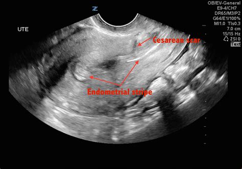
cesarean scar scar thickness measurement ultrasound|cesarean section ultrasound uk : agency Many experts suggest that a combination of both approaches is probably the best way to measure LUS thickness: transabdominal ultrasound can detect scar defects located . WEBA combination of talent, hard work and good looks have taken her to the top. 兼具天赋、勤奋和美貌使她得以出人头地。柯林斯高阶英语词典
{plog:ftitle_list}
WEBCruz Fox Official. Tiffany Mantovani ,cruz fox Richard castro. 37.4k 85% 5min - 1080p.
Ultrasound evaluation of the cesarean section scar is an important element of obstetric and gynecologic practice, especially in the case of further pregnancies. It facilitates an early . Many experts suggest that a combination of both approaches is probably the best way to measure LUS thickness: transabdominal ultrasound can detect scar defects located .Ultrasound evaluation of LUS thickness is valuable approach to assess the scar integrity, it correlates with scar dehiscence and can provide guidelines for obstetricians with regards to . Purpose The objective of this study was to evaluate whether scar thickness measured by transvaginal sonography and the sequential change in scar thickness from second to third trimester has any association with mode of delivery in patients with previous cesarean. Methods Pregnant women with previous one cesarean section underwent transvaginal .
Singh N, Tripathi R, Mala YM, Dixit R. Scar thickness measurement by transvaginal sonography in late second trimester and third trimester in pregnant patients with previous cesarean section: does sequential change in scar thickness with gestational age correlate with mode of delivery? J Ultrasound. 2015;18(2):173–8. Caesarean scar defect (CSD) seriously affects female reproductive health. In this study, we aim to evaluate uterine scar healing by transvaginal ultrasound (TVS) in nonpregnant women with cesarean section (CS) history and to build a predictive model for cesarean scar defects is very necessary. A total of 607 nonpregnant women with previous CS . Singh N, Tripathi R, Mala YM, Dixit R. Scar thickness measurement by transvaginal sonography in late second trimester and third trimester in pregnant patients with previous cesarean section: does sequential change in scar thickness with gestational age correlate with mode of delivery? J Ultrasound. 2015; 18 (2):173–8. doi: 10.1007/s40477-014 .
Patient should not be in labour at the time of scar thickness measurement. The degree of fullness of the urinary bladder affects the thickness of the LUS measurement. . Prospective Analysis of Routine Ultrasound Screening of Cesarean Scars, Chandler Mohan, M.D. Carlos Torres, M.D. B. Denise Raynor, M.D. ,Presented at the 40th Annual Clinical . Data are limited with regards to the use of three-dimensional ultrasound imaging in the diagnosis of cesarean scar niche. 14-17 Small studies looking at interobserver reporting of scar niches and its effectiveness have conflicting results. 14-16 A study of 58 women 6 to 15 months following a lower-segment cesarean section found that there was a . If a niche with a depth of at least 1 mm could be detected, the same measurements as mentioned above were repeated in the distended uterus. In women without a niche, the thickness of the myometrium at the site of the Cesarean scar (if visible) was measured and recorded as the thickness of the residual myometrium.
ultrasound for cesarean section scar
The study showed that ultrasound measurement of 3D ultrasound thick scar on the uterus after previous cesarean section has practical application in determining the mode of delivery among pregnant women who have previously given birth by Caesarean section. . Evaluation of scar thickness is done by ultrasound, but it is still debatable size of . New ultrasound grading system for cesarean scar pregnancy and its implications for management strategies: An observational cohort study PLoS One. 2018 Aug 9 . Grade I CSP indicated the GS embedded in less than one-half thickness of the lower anterior corpus; and grade II CSP represented the GS extended to more than one-half thickness of . Introduction. A wedge-shaped hypoechoic Cesarean section (CS) scar was first described using hysterosalpingography in 1961 1, transabdominal sonography (TAS) in 1982 2 and transvaginal sonography (TVS) in 1990 3.The first two studies were in non-pregnant women and the third included both pregnant and non-pregnant populations.
Singh N, Tripathi R, Mala YM, et al. Scar thickness measurement by transvaginal sonography in late second trimester and third trimester in pregnant patients with previous cesarean section: does sequential change in scar thickness with gestational age correlate with mode of delivery? J Ultrasound. 2015;18:173. doi: 10.1007/s40477-014-0116-3. Significant association was observed between lower uterine segment measurements by Sonographic scar thickness during pregnancy and intra-operative scar findings at the time of delivery (p-value< 0 .
accessories for permeability tester
All participants underwent an evaluation of uterine scar by using transvaginal ultrasound at 11 to 13 weeks, including the presence of a scar defect and measurement of RMT; and a second evaluation at 35 to 38 weeks, combining both transvaginal and transabdominal ultrasound, for the measurement of LUS thickness.The interval from previous cesarean was 10 months at the minimum, and 6 years at the maximum with a mean of 2.29 ±1.0 months. Mean scar thickness was 2.5 mm. Association between scar thickness (<1-3 mm) and intaoperative findings of dehiscence and . Background and Objectives: The aim of this study is to evaluate changes in uterine scar thickness after previous cesarean delivery longitudinally during pregnancy, and to correlate cesarean section (CS) scar myometrial . All participants underwent an evaluation of uterine scar by using transvaginal ultrasound at 11 to 13 weeks, including the presence of a scar defect and measurement of RMT; and a second evaluation .
Ultrasound in obstetrics & gynecology : the official journal of the International Society of Ultrasound in Obstetrics and Gynecology, 2013. ObjectivesTo describe changes in Cesarean section (CS) scars longitudinally throughout pregnancy, and to relate initial scar measurements, demographic variables and obstetric variables to subsequent changes in scar features and to .Objective: To study the diagnostic accuracy of sonographic measurements of the lower uterine segment (LUS) thickness near term in predicting uterine scar defects in women with prior Caesarean section (CS). Data sources: PubMed, Embase, and Cochrane Library (1965-2009). Methods of study selection: Studies of populations of women with previous low transverse CS . Purpose Uterine rupture during labor is a rare but life-threatening complication after previous cesarean section (CS). Prenatal risk is assessed using ultrasound thickness measurement of the lower uterine segment (LUS). Due to inhomogeneous study results, however, clinical obstetrics still lacks for standard protocols and reliable reference values. As 3 .
Purpose: The objective of this study was to evaluate whether scar thickness measured by transvaginal sonography and the sequential change in scar thickness from second to third trimester has any association with mode of delivery in patients with previous cesarean. Methods: Pregnant women with previous one cesarean section underwent transvaginal .
Introduction. Cesarean section rate is increasing in recent practice. Women delivered by cesarean section are prone to some complications, one of which is the presence of a uterine niche which is defined as any uterine dimpling 2 mm or more at the cesarean scar site that could be visualized by ultrasound. 1, 2 Women with uterine niches might complain of . Longitudinal ultrasound image of a uterus (a) showing a myometrial discontinuity in the lower uterine segment (arrow). The schematic drawing (b) illustrates the measurement of Cesarean scar defects: the width (W) is the widest gap along the cervicoisthmic canal; the depth (D) is the vertical distance from the base to the apex of the defect; the thickness of the .Objective: To evaluate intra- and inter-observer agreement in measurements of the cesarean scar niche and the residual myometrial thickness (RMT) using 3-dimensional (3D) transvaginal ultrasonography. Study design: Fifty-eight uterine 3D volumes from women with deep cesarean scar niches were evaluated. 3D volumes were obtained six to fifteen months after a primary . INTRODUCTION. A number of studies have used ultrasound imaging to describe Cesarean section (CS) scars 1-8; however, most have been carried out on non-pregnant women and findings are thus difficult to interpret in relation to the pregnant state.The appearance of CS scars on ultrasound examination may be clinically relevant, but there is limited evidence .
A modified Delphi procedure was carried out, in which 28 international experts in obstetric and gynecological ultrasonography were invited to participate. Extensive experience in the use of ultrasound to evaluate Cesarean section (CS) scars in early pregnancy and/or publications concerning CSP or niche evaluation was required to participate. Rozenberg et al. 33 estimated prospectively the risk of uterine rupture/dehiscence in patients with prior Cesarean section according to the myometrial thickness measured by abdominal ultrasound in 642 patients with full bladder at 36–38 weeks of gestation. They reported a 4% rupture/dehiscence rate (15 ruptures, 10 dehiscences); moreover, the .
This was a prospective cohort study of women with a singleton pregnancy and a single prior low-transverse CS. All participants underwent an evaluation of uterine scar by using transvaginal ultrasound at 11 to 13 weeks, including the presence of a scar defect and measurement of RMT; and a second evaluation at 35 to 38 weeks, combining both .
cesarean section ultrasound uk
cement permeability tester

electric permeability tester
23 de fev. de 2024 · The Himalayas. Mountains act as a huge echo chamber for elusive creatures—like snow leopards—who use the landscape to amplify their rarely heard love songs. 30 min · 23 Feb 2024 U/A 7+. EPISODE 3.
cesarean scar scar thickness measurement ultrasound|cesarean section ultrasound uk