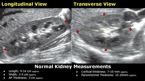
how to measure renal cortical thickness on ultrasound|reduced cortical thickness kidney : factories The cortical thickness is measured from renal capsule to the base of the triangular medullary pyramids. The parenchymal thickness is measured from the renal . WEB149 Followers, 10 Following, 2 Posts - See Instagram photos and videos from LOMOTIFS DE MENINAS (@lomotifs_pesados)
{plog:ftitle_list}
Resultado da 5 dias atrás · ALF is an American sitcom that on Syndication from September 22, 1986, to March 24, 1990, spawning four seasons, 99 episodes and later a television in 1996. It stars an alien named Gordon Shumway, ALF (abbreviation of Alien Life Form), that crash landed on Earth into the .
The cortical thickness is measured from renal capsule to the base of the triangular medullary pyramids. The parenchymal thickness is measured from the renal . Normal kidney appearance in adult 11: cortex is less echogenic than the liver. medullary pyramids are slightly less echogenic than the cortex. cortex thickness equals/is .Key Words: Renal disease, Ultrasound, Renal length, Cortical thickness. Introduction. Measurement of renal size by ultrasound is essential when evaluating patients with possible .Measures of the kidney. L = length. P = parenchymal thickness. C = cortical thickness. Doppler examination of the kidney is widely used, and the vessels are easily depicted by the color .
renal cortical thickness normal range
reduced cortical thickness kidney
The aim of this study was to evaluate the correlations between laboratory findings and ultrasonographic measurements of renal length and cortical thickness in patients .Longitudinal ultrasound image of right kidney shows cortical thickness measured perpendicularly from outer margin of kidney to corticomedullary junction (arrow). Measurement is 0.46 cm.
Key Words: Renal disease, Ultrasound, Renal length, Cortical thickness. Introduction. Measurement of renal size by ultrasound is essential when evaluating patients with possible renal disease. However, it requires prior knowledge .
Over the last 30 years, renal ultrasound (US) has remained the imaging technique of choice to measure renal length in patients presenting with acute or chronic kidney injury, despite the development of alternative .
The renal cortex (parenchyma) is preserved and does not show any atrophy. Editor’s Note: Grade 1 Hydronephrosis (Pelviectasis) can be found in healthy/normal patients that have not voided for a prolonged amount of time. .The normal cortical thickness is 7-10 mm; reduced cortical thickness may indicate progressive kidney disease or decreased eGFR. 11, 15, 16 With respect to volume, the normal range in men is 110-190 mL and in women is 90-150 mL. 17 It is possible to measure volume by simplifying the kidney’s shape as spherical or ellipsoid and measuring .OBJECTIVE. The purpose of our study was to determine whether there is a relationship between renal cortical thickness or length measured on ultrasound and the degree of renal impairment in chronic kidney disease (CKD). MATERIALS AND METHODS. From October to December 2007, 25 patients (13 men and 12 women, mean age 73 years) were identified who had CKD .
OBJECTIVE. The objective of our study was to develop, by use of ultrasound, nomograms of renal parenchymal thickness, medullary pyramid thickness (height), renal length, and the ratio of medullary pyramid thickness to parenchymal thickness in healthy children. SUBJECTS AND METHODS. This prospective study included 292 consecutive children (136 boys and 156 girls) .
Point-of-care (POC) renal ultrasound (US) is a rapid, bedside test for the evaluation of the patient with suspected renal colic or urinary retention. . Grainy gray periphery which is made up of the renal cortex and pyramids. Sometimes you can see the individual pyramids, but this is not always the case. . Measurement of normal right kidney .between renal cortical thickness or length measured on ultrasound and the degree of renal impairment in chronic kidney disease (CKD). MATERIALS AND METHODS. From October to December 2007, 25 patients (13 men and 12 women, mean age 73 years) were identified who had CKD but were not on di-alysis. How To Measure Kidney On Ultrasound | Renal Length, Width, AP Thickness & Volume Measurements USGLength: 9-14 cm approx.Width: 3-5 cm approx.AP Thickness: 3 . The study patients had an average fat-to-cortical thickness ratio <0.4 and had a 63% probability of developing renal failure. The probability increased almost linearly from 65% to 85% for patients with a ratio between 0.4 and 0.5, and then plateaued at 85% probability for a .
The cortical thickness values are 6 to 7 mm on average, according to the measurement scale located on the reproduction where each increment corresponds to 10 mm. (B) Transverse axial section of the superior pole of the left contralateral non-stenotic kidney. The cortical thicknesses were greater than the 8-mm threshold on average, referring to . Cortex The normal renal cortex is uniformly echoic, usually slightly hypo- or isoechoic when compared with the liver parenchyma (right kidney) and hypoechoic when compared with the splenic parenchyma (left kidney). It shows a finely granular echotexture. 7 However, in normal cats, fat accumulation in the cytoplasm of the proximal tubular epithelium . A renal sonogram provides the nephrologist or physician information about renal length, cortical thickness, the echogenicity of the renal cortex, and if pathology is present. . Some nephrologists use the cortical thickness over the renal length measurement to determine the severity of renal failure as well as to follow the patient for .
kidney parenchymal thickness ultrasound
Normal kidney size in adults is closely related to height, but weight, body mass index (BMI), age, and ethnicity can also influence this 4.. Kidney dimensions are typically measured sonographically and used to calculate estimated volume. MRI and CT measurements have been shown to generally correlate well with ultrasonography 5,6.. The mean average .
The renal cortical thickness (RCT) reflects the pathological condition of the kidney, and measuring this parameter can help diagnose renal fibrosis in dogs. The normal reference range of RCT in dogs is broad (3-8 mm) because of the extreme diversity in body size. Therefore, this retrospective, refer .OBJECTIVE. The purpose of our study was to determine whether there is a relationship between renal cortical thickness or length measured on ultrasound and the degree of renal impairment in chronic kidney disease (CKD). MATERIALS AND METHODS. From October to December 2007, 25 patients (13 men and 12 women, mean age 73 years) were identified who had CKD .
Measuring the kidney’s cortical thickness can help detect kidney disease and assess the extent of renal function loss. Renal cortical thickness can be measured using ultrasound imaging techniques, including scanning the kidneys during a transabdominal ultrasound. This method is simple, non-invasive, and cost-effective. .
The widely accepted normal ultrasonographic measurement for kidneys in a cat varies between 3 and 4.3 cm in length. One report proposed that feline kidneys can measure 3.2 to 4.1 cm in length, 2.2 to 2.8 cm in width, and 1.9 to 2.5 cm in height. 1 Currently, there is no widely accepted method for determining ultrasonographically normal kidney size for dogs. To acquire an accurate measurement for the cortex using ultrasound, the operator must have some familiarity with sonographic anatomy of the cortex which appears as isoechoic or sightly hypoechoic relative to the liver and spleen. . The renal cortical thickness was measured in sagittal plane at the level of the mid-kidney. The measurement was . Ultrasound. Ultrasound with Doppler examination of intrarenal vessels is usually performed in patients with CKD, and it is common to have a normal exam. Typical B-mode findings of a long-standing severe CKD (especially stage 5) include 2: reduced renal cortical thickness <6 mm 6 more reliable than length 7. reduced renal lengthbetween renal cortical thickness or length measured on ultrasound and the degree of renal impairment in chronic kidney disease (CKD). MATERIALS AND METHODS. From October to December 2007, 25 patients (13 men and 12 women, mean age 73 years) were identified who had CKD but were not on di-alysis.
The objective of this study was to determine the sonographic measurement of renal cortical thickness (RCT) in adults with normal renal function in Isfahan and its relation with gender, age, height . 2.1. Study Population. This study included 137 patients who underwent abdominal ultrasonography between April 2014 and March 2015. Among them, patients with diabetes mellitus, unilateral kidney, renal tumor, hydronephrosis, solitary cyst greater than 4 cm in diameter, polycystic kidney disease, acute kidney injury defined by risk of renal dysfunction, . A subcostal section, which foreshortens the kidney, often underestimates the length and it is more accurate to measure a coronal or posterior longitudinal section with the beam perpendicular to the renal axis. The cortical thickness of the kidney is generally taken as the distance between the capsule and the margin of the medullary pyramid . In some cases, the kidney may be smaller than normal and have thinning of the outer part of the kidney (called cortex). . Echogenic kidney on ultrasound can sometimes be related to technical factors or normal variation instead of true abnormality. In some cases, fluid in the abdomen (ascites) can make the kidneys look whiter than usual. .
The adult kidney size is variable due to the correlation with body height and age; however, normograms for pediatric kidney size are available. Measure cortical thickness: Cortical thickness should be estimated from the base of the pyramid and is generally 7–10 mm. If the pyramids are difficult to differentiate, the parenchymal thickness can .

WEB25 de jul. de 2022 · 游戏名称: Anemoiapolis:第1章 英文名称: Anemoiapolis: Chapter 1 游戏类型: 冒险解密类 (AVG)游戏 游戏制作: Andrew Quist 游戏发行: Andrew Quist 游戏平台: PC 发售时间: 2022 官方网站: https://store.steampowered.com/app/1522960/Anemoiapolis_Chapter_1/ 游戏介绍 一个 .
how to measure renal cortical thickness on ultrasound|reduced cortical thickness kidney