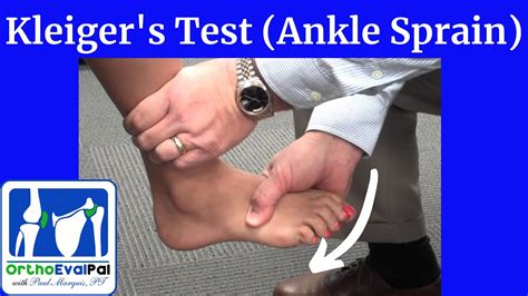
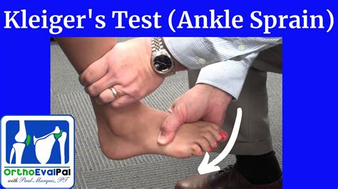
long bone compression test ankle|positive squeeze test foot : consultant Perform the squeeze test just above the anterior tibiofibular ligament. Squeeze the bones together firmly and slowly, hold and then quickly release. If there is pain upon release at the . WEBÉ aqui que entra o “Mais de 9.5 Escanteios”, uma aposta que pode fazer você vibrar como um verdadeiro fã do esporte. Agora preste atenção, porque isso é importante: o .
{plog:ftitle_list}
20 de set. de 2023 · Si su casino HalCash lo tiene como opción de retiro, el proceso es sencillo. Vaya a la sección "cajero" de su casino y seleccione la opción de retirada de HalCash. . 💰 Mejor Casino con promociones de usuarios regulares: 1xbet: 💲 Mejor Casino con Club VIP: LeoVegas: 🤑 Mejor Casino para giros gratis: Megapari: 🎮 Mejor Casino con .
The Long Bone Compression Test is an orthopedic special test utilized in evaluation of suspected foot injury. www.whitworth.edu/msat.Perform the squeeze test just above the anterior tibiofibular ligament. Squeeze the bones together firmly and slowly, hold and then quickly release. If there is pain upon release at the .The examiner stands at the end of the table near the subject's foot. The subject sits with the affected leg extended and the foot off the end of the examining table.The squeeze test compresses the proximal fibula against the tibia to assess the integrity of the bones, interosseus membrane, and syndesmotic ligaments. Pain occurs with fracture or .
Computerized tomography (CT) findings are similar to plain radiographs with sclerosis, new bone formation, periosteal reaction and fracture lines in long bones. It is .
If the diagnosis is uncertain, consider second line investigations including bone scan, computerised tomography or magnetic resonance imaging, and referral to a sports physician. .The following 13 pages are in this category, out of 13 total. Anterior Drawer of the Ankle. Eversion Stress Test. Figure of Eight Method of Measuring Ankle Joint Swelling. Impingement sign .

dr meter moisture sensor meter
Ankle Clearing Test: . First, with the knee bent, ankle is placed in full plantarflexion. The examiner applies compression and scours the joint. Then, with the knee bent, ankle is placed in full dorsiflexion. The examiner applies .In the foot and ankle injury, the use of CT scan is proposed as a modality to assess the passive subsystem. It is a quick tool and it can be used during surgery. A weight-bearing computed tomography (WBCT) allows for the .Purpose: Test for the presence of a stress fracture. Test Position: Sitting or supine. Performing the Test: The examiner places a tuning fork on the suspected site of the stress fracture. Diagnostic Accuracy: Unknown. Importance of Test: . Stress injuries represent a spectrum of injuries ranging from periostitis, caused by inflammation of the periosteum, to a complete stress fracture that includes a full cortical break. They are relatively common overuse injuries in athletes that are caused by repetitive submaximal loading on a bone over time. Stress injuries are often seen in running and jumping athletes .
The seriousness of a broken ankle can vary greatly depending on the type of fracture and the bones involved. The Ankle Joint. The ankle joint is a complex hinge joint where the shinbone (tibia) and calf bone (fibula) meet the talus (talus bone) of the foot. Ligaments hold these bones together and provide stability to the joint.Study with Quizlet and memorize flashcards containing terms like Which of the following should be applied to provide stability for an upper arm injury? A. Traction splint B. Straight arm splint C. Sling and swathe D. Pressure bandage, A primary reason for splinting a bone or joint injury is: A. preventing movement to reduce the chance for further injury. B. setting the bone ends back . Three ligaments keep your ankle bones from shifting out of place. A sprained ankle is when one of these ligaments is stretched too far or torn. . areas where bones grow at the ends of long bones .As a result, the bone weakens and becomes vulnerable to stress fractures. Stress fractures in the foot and ankle occur are most common in the metatarsal bones. They are also often seen in the: Calcaneus (heel) Fibula (the outer bone of the lower leg and ankle) Talus (the lower bone in .
High Ankle Sprain & Syndesmosis Injuries are traumatic injuries that affect the distal tibiofibular ligaments and most commonly occur due to sudden external rotation of the ankle. . compression of tibia and fibula at midcalf level causes pain at syndesmosis. . ossification must be "cold" on bone scintigraphy prior to removal.
Apply 25% tension to the tape and attach the other end of the tape to the outside of your lower leg, covering your ankle bone. Cut two more pieces of tape, long enough to fit around your ankle at the height of your ankle bones. Holding the end of one piece of tape on the inside of your ankle, secure the end to the back of your ankle, at the .
The tarsal tunnel is located inside of the ankle. Running next to the ankle bones, this narrow tunnel is a path for many of the foot and ankle’s tendons, nerves, and blood vessels. . it is usually due to compression or entrapment of the nerve as it passes through the tarsal tunnel. . The test is different than imaging tests like an X-ray . Self-adherent compression bandages, such as Coban or Dynarex, are bandages that behave like tape but do not stick to the skin. They can be torn to specific lengths and come in widths ranging from 1/2 to 4 inches. Self-adherent compression wraps (self-adhesive bandages) are regularly used in athletics or following a blood draw to provide . Bone healing is a natural process. Our bone is constantly being replaced with new bone, and after a bone injury occurs, the body has a tremendous capability to heal the damage to the bone. People who sustain broken bones typically will heal these fractures with appropriate treatment that may include casts, realignment, and surgery.Purpose: To help identify tibiofibular syndesmotic injuries. Test position: Supine. Performing the Test: The examiner grasps the patient's leg midway up the calf and performs a compress and release motion. A positive test is considered if the patient experiences pain in the area of the syndesmosis. Diagnostic Accuracy: Kappa .75. Importance of the Test: The term "high ankle .
squeeze/compression test bump/percussion test long bone compression test morton's test. squeeze/compression test-squeeze the tib/fib together away from fracture site positive finding: . roll the calcaneus outward positive findings: pain, gaping at .
Rest your ankle by not walking on it or returning to sport. Ice should be immediately applied to keep the swelling down. It can be used for 20 to 30 minutes, 3 or 4 times daily. Do not apply ice directly to your skin. .Anterior ankle impingement (footballer's ankle) is a painful pinching or compression of either the soft or the bony tissue at the front of your ankle joint.The calcaneus (heel bone) is the largest of the tarsal bones in the foot. It lies at the back of the foot (hindfoot) below the three bones that make up the ankle joint. These three bones are the: Tibia (shinbone) Fibula (smaller bone in the lower leg) Talus (small foot bone that works as a hinge between the tibia and the fibula)
A simple misstep or twisting injury can cause bone fractures. Treatment depends on the exact site of injury and its severity. . Lightly wrap the injury in a soft bandage that provides slight compression. Request an appointment. By Mayo Clinic Staff. Mar 26, 2022. Print. . Foot and ankle bones. Associated Procedures. Bone scan. CT scan. MRI .
POSITION OF THE ANKLE. STRUCTURES INVOLVED. DESCRIPTION OF TEST BEING PERFORMED. MOUSE OVER PICTURE TO VIEW MOVIE: Eversion Stress. Medial Stress. Neutral plantarflexion to eversion. Deltoid Ligament. Knee is bent 90 0 and gastrocnemius is relaxed. The heel is held from below by one hand while the other hand holds the lower leg. . Acute ankle sprains are commonly seen in both primary care and sports medicine practices as well as emergency departments and can result in significant short-term morbidity, recurrent injuries, and functional instability. Although nonoperative treatment is often successful in achieving satisfactory outcomes, correct diagnosis and treatment is important at the time of .
More than 2 in 5 people with tarsal tunnel syndrome have a history of injuries such as ankle sprains. A sprained ankle is an injury to your ankle ligaments. What are the symptoms of tarsal tunnel syndrome? Tarsal tunnel syndrome causes signs of nerve pain. TTS usually causes pain in the inside of your ankle or the bottom of your feet. Ankle impingement is a syndrome that encompasses a wide range of anterior and posterior joint pathology involving both osseous and soft tissue abnormalities. In this review, the etiology, pathoanatomy, diagnostic workup, and treatment options for both anterior and posterior ankle impingement syndromes are discussed.But pinched or compressed nerves can occur anywhere in the body and the foot or ankle are no exception. When a bone, tendon or ligament presses against a nerve, pain and dysfunction can result. . Make an appointment to treat Nerve Compression in the Foot or Ankle. Call (757) 596-1900 to schedule an appointment with an OSC Physician.
Tarsal Tunnel Syndrome (TTS) is a mononeuropathy caused by compression of the posterior tibial nerve or its branches in the foot/ankle [1]. TTS is analogous to Carpal Tunnel Syndrome, but occurs much more rarely, and usually as a result of trauma (fracture or sprain of the ankle), arthritis, or space-occupying lesions [2].
external rotation stress test, squeeze test and interosseous membrane tenderness length should be performed if the mechanism suggests a syndesmosis injury, or if there is tenderness on palpation of the AitFl (Table 1). importantly, following an ankle inversion plantarflexion injury, 60% of patients will have pain over the medial malleolus in the
A broken ankle can range from a stress fracture to a partial or complete displaced break of the ankle bone. Learn how ankle fractures are diagnosed and treated. > Skip repeated content. . so does the risk of long-term joint damage. Trimalleolar ankle fractures and pilon fractures have the most cartilage injury and, therefore, have a higher .
special tests for ankle sprain

webHours of OPERATION. Sunday 10 am - 10 pm. Monday thru Thursday 4 pm - 10 pm. Friday 4 pm - 1 am. Kitchen closes at 11 pm. Entertainment starts at 9 pm. Saturday 10 am – 1 am. Kitchen closes at 11 pm. Entertainment starts at 9 pm. **Hours are subject to .
long bone compression test ankle|positive squeeze test foot