tear cells blood test|red blood cells teardrop : mfg When evaluating patients with leucoerythroblastic smears (defined by the presence of early myeloid and erythroid forms), the presence of teardrop cells can be helpful in distinguishing often malignant marrow infiltrative . 13 de jul. de 2023 · When looking to begin gambling there are a whole host of places to enjoy the experience. From online casino Canada , games, sports betting to the establishments offered by our high streets. Our guide takes you through the possibilities that come from residing in the province of Newfoundland and Labrador.
{plog:ftitle_list}
webTudo sobre o filme Jogos Vorazes: Em Chamas (The Hunger Games: Catching Fire). Sinopse, trailers, fotos, notícias, curiosidades, cinemas, horários, e muito mais sobre o filme Jogos Vorazes: Em Chamas. . Assistir este filme ontem e agora fica a duvida como sera o terceiro filme sem peeta Nota 17/06/2014 | Responder. Titio Cookies comentou .
what causes tear cells
2000 miata compression test
teardrop blood cells pictures
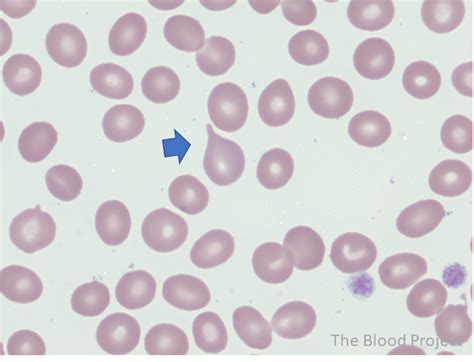
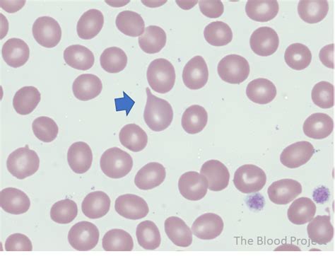
When evaluating patients with leucoerythroblastic smears (defined by the presence of early myeloid and erythroid forms), the presence of teardrop cells can be helpful in distinguishing often malignant marrow infiltrative . Teardrop erythrocytes (syn. dakryocytes) play a key role in the evaluation of peripheral blood smears in patients with anemia, especially as part of the “leukoerythroblastic picture”.Tear Cells (Dacrocytes, Teardrops) – A Laboratory Guide to Clinical Hematology. 17 Tear Cells (Dacrocytes, Teardrops) Michelle To and Valentin Villatoro. Images show peripheral blood .
A dacrocyte (or dacryocyte) is a type of poikilocyte that is shaped like a teardrop (a "teardrop cell"). A marked increase of dacrocytes is known as dacrocytosis. These tear drop cells are found primarily in diseases with bone marrow fibrosis, such as: primary myelofibrosis, myelodysplastic syndromes during the late course of the disease, rare form of acute leukemias and myelophthisis caused by metas. Examination of the peripheral blood smear is an inexpensive but powerful diagnostic tool in both children and adults. In some ways, it is becoming a "lost art," but it often . These abnormal erythrocytes have a teardrop, or pearlike, shape. They are associated with disorders with an abnormal spleen or bone marrow, such as idiopathic myelofibrosis, thalessemia, or myelophthisis. Peter Maslak, .

Images show peripheral blood smears with numerous tear cells (examples indicated by arrows). From MLS Collection, University of Alberta. Image 1: 50x oil immersion.Also known as. Dacrocyte, tailed poikilocyte. Definition. Red cells appearing in the shape of a teardrop or a pear with a single short or long tail. True tear drops have blunted tips and point in different directions. Ddx. Smear artifact. Conditions .
2000 mustang gt compression test
Normal RBCs (also called erythrocytes) are typically disk-shaped, are thinner in the middle than in the edges, with a diameter of 6.2 to 8.2 micrometers, a thickness at the . Teardrop cells in a peripheral blood smear from a patient whose bone marrow was extensively replaced by B lymphoblastic leukemia. Teardrop cells may be seen in the setting of marrow infiltration (by fibrosis, . - Blood smear too thick - Blood smear too thin - ATL peripheral blood smear - Normal blood smear - Rouleaux in myeloma - Blood smear showing RBC agglutination in CAD - Hypertriglyceride milky plasma - Macro-ovalocytes - Elliptocytes (HE) - Schistocytes on peripheral smear - Helmet cells - Waring blender syndrome - Tear drop cells - Spherocytes - Blood .Red blood cells that are teardrop or pear shaped with one blunt projection. 1 The size of these cells are variable. 2. Cell Formation: Red blood cells with inclusions: Teardrop cells form from these cells when the cells attempt to pass through the microcirculation resulting in the pinching the cell as the part containing the inclusion is left .

Teardrop erythrocytes (syn. dakryocytes) play a key role in the evaluation of peripheral blood smears in patients with anemia, especially as part of the “leukoerythroblastic picture”. Teardrop-shaped red blood cells can be seen in a wide range of diseases that lead to bone marrow fibrosis, which is often accompanied by extramedullary hematopoesis. The .Red blood cell, shape abnormality: Teardrop cell: Also known as: Dacrocyte, tailed poikilocyte: Definition: Red cells appearing in the shape of a teardrop or a pear with a single short or long tail. True tear drops have blunted tips and point in different directions. Ddx: Smear artifact: Conditions associated with the shape abnormality No worry: All laboratory results need to be interpreted in the clinical context and the doctor who ordered the tests is usually in the best position to do that. Having said that, it is just the shape of some red cells. A few tear drop cells is no cause for concern, especially if your hemoglobin level is normal.
You can see evidence of the marrow fibrosis and splenomegaly in the blood if you look closely at the red cells. In squeezing through the tight fibrosis in the marrow, and in navigating through a markedly enlarged and cellular spleen, the red cells take on an unusual, “teardrop” shape.
Poikilocytosis is the term used for abnormal-shaped red blood cells (RBCs) in the blood. Normal RBCs (also called erythrocytes) are typically disk-shaped, are thinner in the middle than in the edges, with a diameter of 6.2 to 8.2 micrometers, a thickness at the thickest point of 2 to 2.5 micrometers, and a thickness in the center of 0.8 to 1 micrometers. Poikilocytosis .
Teardrop cells (dacrocytes) are cells which are rounded at one end and tapered at the other. . Peripheral blood film from a patient with myelofibrosis, showing a teardrop cell. . Test Yourself. Beginner Test 1 Morphology Test 1 Morphology Test 2 Return to FRCPath Haematology Part 2: Morphology . LearnHaem | Haematology made simple
Normal red blood cells are round, flattened disks that are thinner in the middle than at the edges. A poikilocyte is an abnormally-shaped red blood cell. [1] Generally, poikilocytosis can refer to an increase in abnormal red blood cells of any shape, where they make up 10% or more of the total population of red blood cells.
Polychromasia occurs on a lab test when some of your red blood cells show up as bluish-gray when they are stained with a particular type of dye. . RDW unable to report due to abnormal histogram, RBC morphology: tear drop cells and Dimorphic RBC population present, normal lymphocytes but high ABN lymphocytes, platelet evaluation: adequate . The peripheral blood smear showed several abnormalities and abnormal cells, with tear drop cells as the most apparent. Teardrop cells are described in a wide range of diseases that are associated . Part of my routine for many years since diagnosed with myeloma has been lab test monthly on average. For the very first time the results indicated a few Tear Drop Cells and a few Ovalocytes. I will look it up enough to formulate sensible questions for my oncologist and for my August Mayo appointment.The presence of teardrop-shaped red cells in peripheral blood has traditionally been felt to reflect altered marrow architecture, namely myelofibrosis. We evaluated two patients with splenomegaly, moderately severe hemolytic anemia due to warm-reactive IgG anti-red cell autoantibody, and bone marrow .
Poikilocytosis is a term that refers to the presence of abnormally shaped red blood cells (RBCs), which are known as poikilocytes. As poikilocytosis is usually a symptom of another medical .
Anisocytosis is the medical term for having red blood cells (RBCs) that are unequal in size. It can be caused by anemia, certain blood diseases, or by some cancer drugs. Normally, a person’s . A test for red blood cell (RBC) count is often part of a complete blood count (CBC) test. In addition to RBCs, this test measures: Hemoglobin (Hb), the protein in RBCs that carries oxygen and carbon dioxide molecules; . A blood smear may also be done to monitor the side effects of chemotherapy or to help diagnose an infection, such as malaria. Normal Results. Red blood cells (RBCs) normally are the same size and color and are a lighter color in the center. The blood smear is considered normal if there is: Normal appearance of cells; Normal white blood cell .
Some red blood cell (RBC) disorders affect the shape of the cells by altering the plasma membrane composition or the ratio of plasma membrane to intracellular volume. Three of the most common morphologies are burr cells (echinocytes), acanthocytes, and target cells. White blood cells can be divided into the myeloid/monocytic cells (neutrophils, eosinophils, basophils, and monocytes) and lymphocytes. Segmented neutrophils are the predominant white cells in the peripheral blood. . differential morphology and LFTs with a couple other tests. She had poikilocytosis 2+, tear cells 1+, burr cells 1+. Her auto .
A recent blood test showed tear cells +1 elliptocytes+1 macrocytes+1 and spherocytes+1. My red cells are 3.8. My hemaglobin is normal. My hematologist said everything looked fine. . My search took me to an interesting site written by a pathology student regarding tear-shaped blood cells. While it mirrors some of the other articles I read on .
How is anisocytosis diagnosed? Healthcare providers may use either of the following tests (or both) to identify anisocytosis. Complete blood count (Red blood cell distribution width): A complete blood count (CBC) is a routine blood test that providers use to check on your blood cells. A particular value on this test called red blood cell distribution .
Teardrop cells are red cells appearing in the shape of a teardrop or a pear with a single, short or long, often blunted or rounded end are called teardrop cells. True tear drops have blunted tips and point in different directions. Teardrop cells are .
Examination of the peripheral blood smear should be considered, along with review of the results of peripheral blood counts and red blood cell indices, an essential component of the initial evaluation of all patients with hematologic disorders. The examination of blood films stained with Wright's stain frequently provides important clues in the diagnosis of anemias and various . Smudge cells are exactly that—cells that were destroyed during the collection or preparing of the blood smear, creating a smudge look on the glass slide. They’re remnants of cells that lack any identifiable cytoplasmic membrane or nuclear structure. Anemia is defined as the reduction in circulating red-cell mass below normal levels. Anemia is a very common condition that is widespread in the human population. Circulating red blood cells (RBCs) contain a protein known as hemoglobin, that protein has four polypeptide chains and one heme ring that contains iron in reduced form. Iron is the main .
Red blood cells are the most abundant blood cells in your body. They make up almost half of your blood's volume. They grow in your bone marrow and take about 7 days to mature.

webDançando pelada mostrando a buceta. 1080p 10 min. Siririca caliente mostrando a buceta. 1080p 6 min. Duas safadas se beijam e ficam peladas, mostrando os peitos e a .
tear cells blood test|red blood cells teardrop