tear film function tests|biochemistry tear film : Brand Assessment of the tear breakup time, Schirmer test, and lipid layer allows detection of the type of tear film deficit. Evaluating the presence of ocular surface inflammation, extent and location of staining, and other adnexal conditions . The famous Akinator has come to Miniplay for you to try to beat him in his own game. The genius will ask you up to 25 questions to try and guess the character, real or fictional, or the animal you have in mind.
{plog:ftitle_list}
Casa a Venda Na Ilha Composta de 6 Suítes, Sendo 3 Suítes no Pavimento Superior e 3 Suítes no Pavimento Térreo, o Terreno Possuí 1600 m² É um verdadeiro quadro "Vivo" ! .

the tear film
This chapter reviews the current understanding of the structure of the tear film and will introduce the concept of the integrated lacrimal functional unit as a key component of the healthy ocular . It follows that assessing the quantitative and qualitative status of the tear film using the TMH, RBS, and TBUT tests is important for detecting tear film dysfunction, and that . This study aims to evaluate the impact of tear film instability on visual function and determine the effectiveness of PBBT as an alternative option for assessing tear film .This review examines various techniques that are used to assess tear film instability: evaluation of tear break-up time and non-invasive break-time; topographic and interferometric techniques; .
Assessment of the tear breakup time, Schirmer test, and lipid layer allows detection of the type of tear film deficit. Evaluating the presence of ocular surface inflammation, extent and location of staining, and other adnexal conditions . This review examines various techniques that are used to assess tear film instability: evaluation of tear break-up time and non-invasive break-time; topographic and . Abstract. The precorneal tear film is a thin layer, about 2–5.5 μm thick, which overlays the corneal and conjunctival epithelium. It functions to lubricate and protect the corneal and eyelid interface from environmental .
Tear production is approximately 1.2 microliters per minute, with a total volume of 6 microliters and a turnover rate of 16% per minute. 2 Tear film thickness, as measured by interferometry, is 6.0 µm ± 2.4 µm in normal subjects and is significantly thinner in dry eye patients with measured values as low as 2.0 µm ± 1.5 µm . 3Tear film stability can be assessed via a number of tools designed for clinical as well as research purposes. . -invasive break-time; topographic and interferometric techniques; confocal microscopic methods; aberrometry; and visual function tests. It also describes possible contributions of different tear film components; namely meibomian .Tear film function tests such as tear break up time (TBUT), tear meniscus height (TMH), and Schirmer's I and II tests were conducted. For dry eye subjects, salivation was induced by giving a tamarind candy (a soft slightly sour tamarind pulp mixed with sugar) for 5 min. Tear film function tests were carried out within a few seconds (2 to 3 s . Tear film function tests such as tear break up time (TBUT), tear meniscus height (TMH), and Schirmer’s I and II tests were conducted. For dry eye subjects, salivation was induced by giving a tamarind candy (a soft slightly sour tamarind pulp mixed with sugar) for 5 min. Tear film function tests were carried out within a few seconds (2 to 3 s .
A TBUT test checks how long your tear film (layer of tears on your eyes) lasts after you blink. Your eye doctor will place a small amount of dye in your eye, and you’ll blink to make the dye fully cover your eye. Then, you’ll look forward without moving your eyes or blinking. Your eye doctor will watch to see how long the dyed tear film .component of the healthy ocular surface. Currently available techniques to evaluate tear film function and stability are discussed. Finally, current theories of dry eye disease are reviewed to add insight into the biology of the normal tear film. TEAR FILM STRUCTURE The tear film is responsible for providing a smooth refractive surface for . These tests included fluorescein staining, rose Bengal staining, PRT, Schirmer, tear meniscus height and tear film break-up time (TBUT). These finding highlight the poor correlation that exists between all the tests as well as the difficulty in selecting entry criteria for a therapeutic drug study in the dry eye patient population.
Schirmer test with anesthetic to measure basal tear production. Fluorescein staining to test for tear film breakup time (TBUT), which is defined as the interval between the last complete blink and the first appearance of a dry spot or disruption in the tear film. Once in the tear film, fluorescein should stay intact for at least 10 seconds . Investigating whether diabetes mellitus is correlated with tear film dysfunction and/or tear hyposecretion found reflex tearing was demonstrated to be significantly decreased in insulin dependent diabetics and unstimulated basal tear flow and tear film break up time were found to be normal. BACKGROUND—Diabetic patients often complain of dry eye symptoms, .
Tear film tests such as Schirmer's test and tear film breakup time test were performed on all these eyes. Results of tear function test between the eyes with pterygium (cases) and the eye without .
Objective: To evaluate the diagnostic performance of the tests included in primary Sjogren's syndrome (SS-I) diagnostic criteria (Schirmer I, break-up time, vital dye staining) and to compare them with other examinations related to the ocular surface status. Methods: Clinical and cytological data were collected from 177 patients (62 SS-1, 56 non-SS autoimmune diseases, .

Therefore, current methods used to assess tear film anatomy and function should focus on these key factors. Ideally, these methods should be simple and easy to use in every ophthalmic clinic. . Conventional methods such as Schirmer test, fluorescein tear breakup time, and assessment of the quantum and quality of the meibum secretions by .
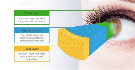
3 Layers of the Tear Film. The tear film comprises 3 distinct layers, each with a specific function: the lipid, aqueous, and mucin layers.. The lipid layer is the outermost layer and consists of a thin film of oil that helps to prevent evaporation of the aqueous layer and keeps the tear film stable and smooth.; The aqueous layer is the middle layer and is composed primarily .The precorneal tear film was first described by Wolff in 1946 as a three-layered structure composed of a superficial lipid layer, a middle aqueous layer, and an deeper mucin layer.[] A more recently proposed model suggests a two-layered structure for the tear film with the superficial lipid layer protecting a hydrated mucogel. Membrane-associated mucins (MUC1, 4, .Purpose: To compare histologic abnormalities of tear film and tear osmolarity between normal eyes and eyes with pterygium. Methods: This was a prospective, hospital-based, case-control study involving 95 patients (65 men, 30 women) with unilateral pterygium. The tear meniscus height (TMH), Schirmer's test-1 (SCH-1) score, Rose Bengal staining (RBS) score, tear film . The DEWS identified increased tear osmolarity and tear film instability as “core mechanisms” of DED, regardless of the etiology. 2 Tests that accurately measure tear osmolarity and tear film instability should therefore, theoretically be best for identifying and determining the severity of DED. Tear osmolarity is attractive, because it .
2. PREFACE • The main aspect of this project is to show the different tear function test to show any abnormality in the tear film or tear film dysfunction. • This give us a brief idea about the abnormality and proper care .
tear film levels
Dr. Monika Soni presented on the topic of tear film at the upgraded department of ophthalmology at MGMMC & MYH Indore. The presentation discussed the anatomy and physiology of tear film, including the three layers of the tear film, mechanisms of tear secretion and distribution, functions of the tear film, tests to evaluate tear film such as tear breakup .However, most people with dry eye syndrome/tear film dysfunction have signs of it which are not even obvious on a general, screening eye exam. Depending on the particular constellation of signs, symptoms, history and comorbidities, tests ranging from Schirmer's tear test to blood tests to check for systemic disease may be warranted.
Some are easily accessible to clinicians, while others remain in the field of clinical research. All of these tests provide a better understanding of the pathophysiology of the tear film. This review hopes to provide an overview of the existing tests and their role in evaluating the significance of the tear film in visual function. Tear film evaluation was done through Tear film Break Up Time (T-BUT) and Schirmer’s Test in patients preoperatively and postoperatively on day 1, 4 weeks, and 8 weeks follow up. CTRI Registration: REF/2023/07/071130 (30/07/2023) Protocol version : .(21.8 7.37) (P 0.01, Student’s t test). The abnormality in each tear test, except those for meibomian gland function and tear meniscus height, was significantly related to the H-Y scores (P 0.05, 2 linear-by-linear association). Conclusion: The results of this study indicate that PD is associated with disturbances in tear function. With Background The tear film breakup time (tBUT) is a clinical evaluation of evaporative dry eye disease assessed by instilling topical fluorescein into the eyes. In the present study, we introduce a new diagnostic test, blinking tolerance time (BTT), for self-evaluation of tear-film stability. We compared the results with the tBUT and validated the BTT test for self .
Tear film tests such as Schirmer's test and tear film breakup time test were performed on all these eyes. Results of tear function test between the eyes with pterygium (cases) and the eye without .The ocular surface requires a complete tear film to maintain its health and function; adequate production, retention, and balanced elimination of tears is necessary for this process. . The advent of new technology, making clinical testing of this feature of tear physiology simple, practical, and inexpensive could provide an impetus to its . Citation 2 Tests that accurately measure tear osmolarity and tear film instability should therefore, theoretically be best for identifying and determining the severity of DED. Tear osmolarity is attractive, because it offers an objective numerical output that can be monitored; other commonly used diagnostic tests rely heavily on subjective .
Measurement of tear-film osmolarity may be performed to assess patients suspected of having dry eye disease, an application probably first considered and promoted by Gilbard et al. Tear-film osmolarity has been shown to be elevated in patients with dry eyes. It is a very sensitive test for identifying a dry eye but lacks some specificity in .

what is moisture meter
Sonho meu, sonho meu Vai buscar quem mora longe, sonho .
tear film function tests|biochemistry tear film