test for retinal tear|does a retinal tear hurt : solution Your healthcare team may use the following tests and instruments to diagnose retinal detachment: Retinal exam. Your healthcare professional may use an instrument with . Resultado da 7 de mai. de 2021 · LiveLeak, a website best known for hosting violence and gory footage that mainstream sites wouldn’t touch, has shut down after .
{plog:ftitle_list}
Resultado da Мы объединение двух даберов - Yupi и Nata_Kex (+ Gin), занимаемся озвучкой и адаптацией иностранного контента.
signs of a torn retina
The Amsler Grid test can be an important indicator of diseases in the retina. Test your eyes daily to detect changes as early as possible. Your healthcare team may use the following tests and instruments to diagnose retinal detachment: Retinal exam. Your healthcare professional may use an instrument with .
However, prompt treatment is needed. A retinal tear can quickly progress to a retinal detachment, which can cause permanent vision loss. This article discusses retinal tears. It examines the symptoms, causes, and .
iphone se apple leather case drop test
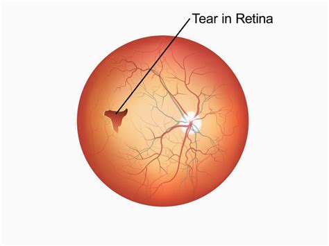
An eye doctor can check for a retinal detachment or retinal tears with a dilated eye exam. During this test, they give you eye drops to widen your pupil and examine the back of your eye. A retina specialist will check for retinal tears by placing drops in your eyes to dilate the pupil. They’ll look through a special lens to assess any changes inside the eye. Eye surgery is the most common treatment for a torn . This test uses high-frequency sound waves, called ultrasonography, to help view the retina and other structures in the eye. It also can identify certain tissue characteristics that can help in the diagnosis and . A retinal tear is a rip in the layer of light-detecting cells at the back of your eye called the retina. It’s a medical emergency that can lead to permanent vision loss if not treated.
A thorough and timely examination by a retina specialist using scleral depression (applying slight pressure to the eye) and/or a 3-mirror lens is the most important step in diagnosing a .The Amsler grid is an at-home test that can help detect changes in the central vision. It is helpful in following the progress of any disease in the macula, the part of the eye responsible for central vision. This is a commonly used test to .
Retinal tears are typically treated with laser or a freezing procedure (cryotherapy). Treatment is performed in an office setting and is very effective and quite safe. Topical or local anesthesia is utilized, and the procedure is only mildly uncomfortable. The treatment creates spot-welding around the edges of the tear that nearly eliminates . A dilated eye exam can help your eye doctor find a small retinal tear or detachment early, before it starts to affect your vision. . a dilated eye exam, you may get an ultrasound or an optical coherence tomography (OCT) .
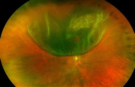
Having a retinal tear or detachment in the other eye; Having family members with retinal detachment; Having weak areas in the retina (which your ophthalmologist may see during an exam) Early Signs of a Retinal Tear. A torn retina has to be checked by an ophthalmologist right away. Otherwise, your retina could detach and you could lose vision in . had a retinal tear or detachment in your other eye; have family members who had retinal detachment; have weak areas in your retina (seen by an eye doctor during an exam) Early Signs of a Detached Retina. A detached retina has to be examined by an ophthalmologist right away. Otherwise, you could lose vision in that eye.Dr. Khan: If a tear develops in the retina, fluid can get in underneath that tear and just lift the retina off like wallpaper off a wall and that's a retinal detachment. Mr. Howland: And that can cause blindness, which is why it's especially important to have a dilated eye exam within days of noticing new floaters or changes in vision. Retinal tears may occur as people age when their vitreous shrinks and pulls the retina from the back of the eye. If a tear occurs, a person may notice a sudden increase in floaters, dark spots in .
A small tear in your retina lets the gel-like fluid called vitreous humor travel through the tear and collect behind your retina. The fluid pushes the retina away, detaching it from the back of your eye. . This imaging test combines X-rays with a computer and is usually used if there’s a history of trauma or possible penetrating eye injury .However, a sudden increase of floaters, which might look like a shower or a curtain of floaters, signifies a retinal tear or detachment. Other warning signs of a retinal tear include flashes of light, shadows or veils over your vision, sudden blurry vision, or decreased peripheral vision. If you notice any of these symptoms, call us right away .
A retinal tear is a rip in the layer of tissue in the back of your eye called the retina. A retinal tear can lead to permanent vision loss, but most people who promptly get medical attention heal .
Causes. In general, retinal detachments can be categorized based on the cause of the detachment: rhegmatogenous, tractional, or exudative. Rhegmatogenous (reg ma TODGE uh nus) retinal detachments are the most common type. They are caused by a hole or tear in the retina that allows fluid to pass through and collect underneath the retina, detaching it from its .
The cause of retinal detachment depends on the type you have: Rhegmatogenous. These are the most common type. Rhegmatogenous detachments result from a hole or tear in the retina. A retinal tear allows fluid to pass into and collect beneath the retina. Gradually, the retina pulls away from the back of the eye, causing blood loss and decreased . Doctors can diagnose a retinal tear by applying slight pressure to the eye during a test called a scleral depression, or by using a special mirror lens. Sometimes, doctors may use what is called an ophthalmic ultrasound if the retina is obscured due to a hemorrhage.Retinal tears are typically treated with laser or a freezing procedure (cryotherapy). Treatment is performed in an office setting and is very effective and quite safe. Topical or local anesthesia is utilized, and the procedure is only mildly uncomfortable. The treatment creates spot-welding around the edges of the tear that nearly eliminates .The most common types of retinal hemorrhages include: Intraretinal hemorrhages: Intraretinal hemorrhages are tears or breaks in the tissue of your retina itself.They’re sometimes called dot-and-blot hemorrhages. Retinal nerve fiber layer hemorrhages: The retinal nerve fiber layer (RNFL) is the group of nerve cells that connect your retina to your optic nerve.

Retinal imaging is a relatively new eye test that can detect many diseases in the eye. WedMD explains what the test is. . Tear Duct Problems; Vitreous Humor Problems; Common Vision Problems . Retinal tears may occur without symptoms, but often photopsia (luminous rays or light flashes in vision) is noted. Photopsia results from mechanical stimulation of the retina by vitreoretinal .The test is usually painless, other than maybe feeling a little pressure or a slight sting. . If you experience symptoms in the other eye, you’ll need a repeat eye exam to be sure there isn’t a retinal tear or a detached vitreous in your other eye. Most people don’t develop complications like a retinal tear. But you should have an eye .
Narrator: An Amsler grid is a simple at home test us to monitor vision. This test is used to assess the macula, the center of the retina, responsible for detailed central vision. The Amsler grid consists of evenly spaced horizontal and vertical lines printed on white paper. A small dot is located in the center of the grid for fixation.Retinal Tears and Holes. Retinal breaks are defined as any full thickness tears or holes in the retina. They are key risk factors for retinal detachments. Retinal tears are usually caused by traction on the retina from a PVD. Retinal holes are commonly caused by vitreous traction or lattice degeneration creating an atrophic hole. Symptoms
A retinal tear is a small split or hole in the lining of the back of the eye. Read about its causes, symptoms, diagnosis, and why timely treatment is important.These routine vision tests don't find retinal detachment itself. But they can find problems that could lead to or result from retinal detachment. A doctor can usually see a retinal tear or detachment while checking the retina using ophthalmoscopy. This test allows the doctor to see inside the back of the eye using a magnifying tool with a light.
Clinical examination alone is usually sufficient to diagnose a retinal tear or detachment. Laboratory tests are typically not necessary. An ultrasound is needed to identify possible retinal tears and/or detachment when vitreous hemorrhage prevents direct visualization of the retina although there has been variable sensitivity (44-100%) in .While retinal tears may occur as a result of eye trauma, most retinal tears occur spontaneously due to a PVD. Another cause of a retinal hole or tear is thinning or weakening of the tissues of the retina over time. Is there a difference between retinal holes and retinal tears? The terms retinal hole and tears may often be used interchangeably .
Cutting-Edge Diagnostics. Early detection of retinal detachment or tear is crucial in order to preserve your eyesight. The best-in-class retinal detachment doctors at VRMNY use all types of ophthalmic imaging, including fluorescein angiography and indocyanine green angiography, as well as noninvasive imaging techniques like spectral domain optical .
Retinal detachment is a serious eye condition that happens when your retina pulls away from the tissue around it. Learn more about the types, causes, risk factors, symptoms, diagnosis, treatment . Boldrey et al. found a higher fellow eye retinal tear rate of 2.6% , but the patients were referred from a retinal practice and the overall retinal tear rate was 18.5% which may represent patient .A tear or hole in the retina (this is the most common cause) A fluid build-up under the retina caused by an eye injury or trauma; Scar tissue within the eye that can pull the retina; Risk factors of retinal detachment. Retinal detachment can happen at any age, but it’s more likely to occur in people who: Are over 40; Are very short sighted

iphone se drop test 1000 feet
15 de jan. de 2024 · Para entrar em contato com a PlayPix, o cliente deve utilizar o e-mail ou o chat ao vivo. Os canais de comunicação são de fácil acesso, podendo ser acessados tanto no computador quanto no celular. A seguir, você confere mais detalhes sobre o suporte da PlayPix: Chat ao vivo: disponível em português, de segunda à sábado, das .
test for retinal tear|does a retinal tear hurt