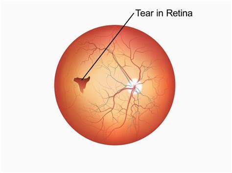
test ofr retinal tear|is a retinal tear serious : distribution If you have a retinal tear, you must seek prompt treatment to prevent retinal detachment and vision loss. Common symptoms of a torn retina include a sudden appearance of floaters or flashes of light. Retinal tear .
WEBO São Paulo inscreveu James Rodríguez no Campeonato Paulista nesta segunda-feira (26). Conforme regulamento, o meio-campista colombiano ficou com. Leia agora. 1 2 3 . » Última ». categorias. Basquete. Futebol. Futebol de Base. Futebol Feminino.
{plog:ftitle_list}
Resultado da Onde assistir ao jogo entre Racing x Newell’s Old Boys? A partida terá transmissão ao vivo pelo serviço de streaming do Star+ e pela ESPN 4 na TV fechada. Racing. Eliminado também da Copa Argentina, o Racing vai lutando para recuperar o foco na Copa da Liga Profissional Argentina, sua única .
The Amsler Grid test can be an important indicator of diseases in the retina. Test your eyes daily to detect changes as early as possible. If you normally wear reading glasses, please wear them while performing this test. Proper lighting . Retinal tears. When the retina has a tear or hole but hasn't yet become detached, your eye surgeon may suggest one of the following treatments. These treatments can help . A retinal tear can cause you to see stars, flashes of light, and specks or squiggles floating across your eye. The retina is a light-sensitive part of the eye that transmits images to the brain. Most retinal tears are caused by . A retinal tear is a rip in the layer of light-detecting cells at the back of your eye called the retina. It’s a medical emergency that can lead to permanent vision loss if not treated quickly..
A thorough and timely examination by a retina specialist using scleral depression (applying slight pressure to the eye) and/or a 3-mirror lens is the most important step in diagnosing a .
If you have a retinal tear, you must seek prompt treatment to prevent retinal detachment and vision loss. Common symptoms of a torn retina include a sudden appearance of floaters or flashes of light. Retinal tear . Diagnosis: Dilated eye exam. Treatment: Laser treatment, surgery. If you have symptoms of retinal detachment, go to your eye doctor or the emergency room right away. Retinal detachment can cause permanent vision . Diagnosis. Treatment. Prognosis. Complications. When to See a Doctor. FAQs. While usually easy to treat, retinal tears can cause retinal detachment if left untreated, which . Retinal tears may occur as people age when their vitreous shrinks and pulls the retina from the back of the eye. If a tear occurs, a person may notice a sudden increase in floaters, dark spots in .

A dilated eye exam can help your eye doctor find a small retinal tear or detachment early, before it starts to affect your vision. . a dilated eye exam, you may get an ultrasound or an optical coherence tomography (OCT) . had a retinal tear or detachment in your other eye; have family members who had retinal detachment; have weak areas in your retina (seen by an eye doctor during an exam) Early Signs of a Detached Retina. A detached retina has to be examined by an ophthalmologist right away. Otherwise, you could lose vision in that eye.How is an Amsler test done? Test each eye on a daily basis to detect early changes in vision. Use the Amsler grid in the same way each time you test your vision. If you normally wear reading glasses, please wear them while performing this test. Good lighting is essential. 1) Hold the Amsler grid at a normal reading distance. 2) Test each eye . Retinal tear. A retinal tear occurs when the clear, gel-like substance in the center of your eye, called vitreous, shrinks and tugs on the thin layer of tissue lining the back of your eye, called the retina. This can cause a tear in the retinal tissue. It's often accompanied by the sudden onset of symptoms such as floaters and flashing lights.
testing bottled water purity
A retinal tear is a rip in the layer of tissue in the back of your eye called the retina. A retinal tear can lead to permanent vision loss, but most people who promptly get medical attention heal .If you have severe myopia (nearsightedness) or have had eye surgery or an eye injury, you have a higher chance of having a retinal tear. Retinal tears deprive your retina of oxygen, which can lead to permanent damage and vision loss. However, the small tear can also allow liquid to seep under the retina, which causes detachment.As the vitreous separates or peels off the retina, it may tug on the retina with enough force to create a tear. Most of the time it doesn't. But if a PVD causes a tear and the tear isn't treated, the liquid vitreous can pass through the tear into the space behind the retina. This causes the retina to .
The cause of retinal detachment depends on the type you have: Rhegmatogenous. These are the most common type. Rhegmatogenous detachments result from a hole or tear in the retina. A retinal tear allows fluid to pass into and collect beneath the retina. Gradually, the retina pulls away from the back of the eye, causing blood loss and decreased . Retinal tears may occur without symptoms, but often photopsia (luminous rays or light flashes in vision) is noted. Photopsia results from mechanical stimulation of the retina by vitreoretinal .Causes. In general, retinal detachments can be categorized based on the cause of the detachment: rhegmatogenous, tractional, or exudative. Rhegmatogenous (reg ma TODGE uh nus) retinal detachments are the most common type. They are caused by a hole or tear in the retina that allows fluid to pass through and collect underneath the retina, detaching it from its .
Key takeaways: A retinal tear is a break or hole in the retina, the part of the eye that sends light signals to the brain. People who are nearsighted, over the age of 60, or had cataract surgery are more likely to develop retinal tears.
A small tear in your retina lets the gel-like fluid called vitreous humor travel through the tear and collect behind your retina. The fluid pushes the retina away, detaching it from the back of your eye. . This imaging test combines X-rays with a computer and is usually used if there’s a history of trauma or possible penetrating eye injury .Retinal tears are typically treated with laser or a freezing procedure (cryotherapy). Treatment is performed in an office setting and is very effective and quite safe. Topical or local anesthesia is utilized, and the procedure is only mildly uncomfortable. The treatment creates spot-welding around the edges of the tear that nearly eliminates . Retinal imaging takes a digital picture of the back of your eye.It shows the retina (where light and images hit), the optic disc (a spot on the retina that holds the optic nerve, which sends .A tear or hole in the retina (this is the most common cause) A fluid build-up under the retina caused by an eye injury or trauma; Scar tissue within the eye that can pull the retina; Risk factors of retinal detachment. Retinal detachment can happen at any age, but it’s more likely to occur in people who: Are over 40; Are very short sighted
Retinal Tears and Holes. Retinal breaks are defined as any full thickness tears or holes in the retina. They are key risk factors for retinal detachments. Retinal tears are usually caused by traction on the retina from a PVD. Retinal holes . Macular degeneration affects the macula, a small part of the retina in the back of your eye. It’s the leading cause of blindness in older adults. About 11 million Americans of all ages are .Narrator: An Amsler grid is a simple at home test us to monitor vision. This test is used to assess the macula, the center of the retina, responsible for detailed central vision. The Amsler grid consists of evenly spaced horizontal and vertical lines printed on white paper. A small dot is located in the center of the grid for fixation.
signs of a torn retina
retinal tear detachment
With YAG vitreolysis, there is a risk of glaucoma, retinal tear, retinal detachment, cataract if you hit the lens, and retinal damage if you hit the retina, said Dr. Chirag Shah. To minimize risks of lens or retinal damage, he recommends ensuring a safe distance between the focal point of the laser and the retina and crystalline lens. Retinal tears occur when the vitreous gel inside the eye pulls away from the retina, causing a tear or hole in the delicate tissue. This can lead to a variety of symptoms, including floaters, flashes of light, and a sudden decrease in vision. . additional tests or imaging may be required to assess the extent of the retinal tear and plan for . Doctors can diagnose a retinal tear by applying slight pressure to the eye during a test called a scleral depression, or by using a special mirror lens. Sometimes, doctors may use what is called an ophthalmic ultrasound if the retina is obscured due to a hemorrhage.
Tears in the retina. Prior history of retinal detachment. Prior eye surgery. Eye trauma. Infection. Diabetes-related retina disease. Eye inflammation. . They may also use the following tests to diagnose macular pucker: Amsler grid eye test, which checks for distorted vision using a page of small squares formed by horizontal and vertical lines.The test is usually painless, other than maybe feeling a little pressure or a slight sting. . If you experience symptoms in the other eye, you’ll need a repeat eye exam to be sure there isn’t a retinal tear or a detached vitreous in your other eye. Most people don’t develop complications like a retinal tear. But you should have an eye .
In addition to the eye exam, imaging tests may also be used to diagnose a retinal tear. Optical coherence tomography (OCT) is a non-invasive imaging test that uses light waves to create detailed cross-sectional images of the retina. . Retinal tear surgery is a procedure that repairs a tear or hole in the retina, the thin layer of tissue at . Retinal tears need to be treated in a timely manner and with proper treatment, otherwise, they can have serious impacts on your long-term vision and can lead to a more serious condition called retinal detachment. A retinal tear can safely be treated with laser surgery. About Retinal Tears. Retinal tears are typically painless.
In our nationwide eye exam cost survey, we found that eye doctors charged an average range of to for a digital retinal test. Retinal imaging tests and devices. There are many ways an eye doctor can examine your retinas — and take their photos. These imaging tests and devices may include: Optomap. One of the most common retinal .
testing bottled water science fair project
Jogos de panelas baratos. economiza frete Em carrinhos de compras. Ordenar por. Mais relevantes. Jogo De Panelas Alumínio Teflon 8 Pc Tampa De Vidro Barato. R$ 189 90. .
test ofr retinal tear|is a retinal tear serious