tests for ptfl tear|Posterior talofibular ligament : exporters The PTFL is an intracapsular but extra-synovial ligament that arises from the posterior aspect of the distal fibula and courses posteromedially to insert into the lateral . Resultado da 8 de dez. de 2022 · bet365 are thrilled to announce the launch of Mercenary X, an exclusive addition to the brand’s portfolio of innovative and cutting .
{plog:ftitle_list}
19 de fev. de 2024 · Por GIGA-SENA. O sorteio do concurso 3032 ocorreu no dia 19 de fevereiro de 2024 e o prêmio principal foi estimado em R$ 1.700.000,00 (um milhão e setecentos mil reais) para quem acertar o resultado da Lotofácil 3032. Quem acertar 14 (quatorze), 13 (treze), 12 (doze) ou 11 (onze) números também ganha prêmio de menor .
AP and mortise ankle radiographs. used to evaluate the tibiofibular clear space and tibiofibular overlap. tibiofibular clear space should be < 5 mm. tibiofibular overlap for AP view > 10 mm. weight bearing mortise view is most . Ankle sprains involve an injury to the ATFL and CFL and are the most common reason for missed athletic participation. Treatment usually includes a period of immobilization followed by physical therapy. Only when .
Indications for imaging studies in cases of suspected talofibular ligament injuries include the following: Bony tenderness or deformity. Suspicion of a fracture or syndesmotic injury. Severe . History. The history portion of the examination for a suspected talofibular ligament injury should include the following: Mechanism of injury. Time of injury. Concurrent injuries. .
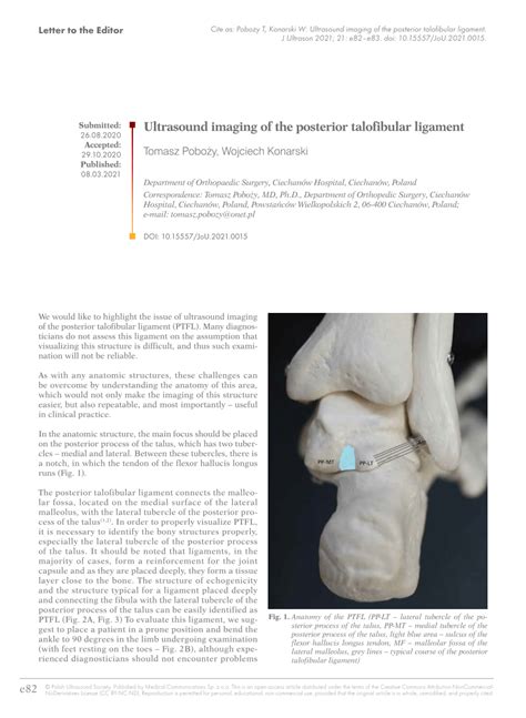
The PTFL is an intracapsular but extra-synovial ligament that arises from the posterior aspect of the distal fibula and courses posteromedially to insert into the lateral .Squeeze Test or Thompson’s Test. To confirm a suspected Achilles tendon rupture, have the patient lying prone and then squeeze the calf while observing the foot. If there is plantar flexion of the foot, this means that the tendon is intact.Posterior talofibular ligament injury involves damage to the ligament that stabilizes the ankle joint, often resulting from trauma or excessive stress. In order to properly visualize PTFL, it is necessary to identify the bony structures properly, especially the lateral tubercle of the posterior process of the talus. It should be noted .
“MRI was able to accurately diagnose lateral ankle ligament tears in most cases. Diagnosis of a complete ATFL tear on MRI is more sensitive than that of complete CFL tear. .
Anterior Drawer Test. Assesses: Anterior talofibular ligament (ATFL) Position: Knee joint in flexion and ankle in 10-15 degrees plantar flexion Maneuver: The examiner exerts a downward force on the tibia while .
However, an asymmetrical thickening and partial tear of the PTFL can occur anywhere. Therefore, measurement mistakes could occur in some cases. In contrast to the PTFL thickness (PTFLT) between anterior and posterior fiber, the cross-sectional area of PTFL does not worry about these measurement mistakes because the PTFL cross-sectional area .Discover the essentials of Anterior Talofibular Ligament (ATFL) tears, a common ankle injury among athletes and active individuals. Understand symptoms, treatments, and recovery timelines to ensure effective healing. Learn about the R.I.C.E. method for immediate care, the importance of early diagnosis, and the role of physical therapy in rehabilitation. Prevent future sprains with .The Talar Tilt Test is a common orthopedic tests to be performed after an inversion trauma in order to assess the ankle ligaments. Skip to content . And lastly, to put the most stress on the posterior talofibular ligament (pTFL), bring the foot into maximal dorsiflexion and perform the same movement again.Figure 4. Lateral talar tilt test Table 1. Clinical testing for syndesmosis injury External rotation stress test The patient’s ankle is passively dorsiflexed in maximal external rotation (either seated or lying prone with knee flexed to 90 degrees). Pain at the syndesmosis is regarded as a positive test Squeeze test With both hands clasp the .
The PTFL originates from the posteromedial aspect of the distal fibula and courses medially to insert onto the lateral aspect of the posterior talus. . but are not limited to, peroneal tendon tears; chondral and osteochondral fractures of the talus; medial ligamentous injury; ankle syndesmosis injury; and fractures to the hindfoot, midfoot .(PTFL), calcaneofibular ligament (CF) are responsible for resistance against inversion . • Tendon tears or tendonitis – current or past . Review any diagnostic imaging, tests, work up and operative report listed under LMR. History of Present Illness: Interview patient at time of examination and include onset, acute, Instability with these tests indicates a complete tear of the ATFL and at least a partial tear of the CFL. Perform a neurologic exam. This should include testing the patient's balance. Have them stand on their uninjured foot, initially with their eyes open; then, have them close their eyes. Then have the patient do this with the injured foot . ss. Thus, we created the PTFL cross-sectional area (PTFLCSA) as a diagnostic image parameter to assess the hypertrophy of the whole PTFL. We assumed that the PTFLCSA is a key morphological diagnostic parameter in CLAI. PTFL data were obtained from 15 subjects with CLAI and from 16 normal individuals. The T1-weighted axial ankle-MR (A-MR) images .
Ankle sprains are a common reason for presentation to the emergency department, accounting for approximately 7% to 10% of visits and up to 40% of all sports injuries.[1] The majority of ankle injuries are sports-related and involve the lateral ankle compartment. The lateral ankle ligaments consist of the anterior talofibular ligament (ATFL), the calcaneofibular ligament . The PTFL originates from the posteromedial aspect of the distal fibula and courses medially to insert onto the lateral aspect of the posterior talus. . but are not limited to, peroneal tendon tears; chondral and osteochondral fractures of the talus; medial ligamentous injury; ankle syndesmosis injury; and fractures to the hindfoot, midfoot .The CFL and the PTFL can also be injured and, after severe inversion, subtalar joint ligaments are also affected. Commonly, an athlete with a lateral ankle ligament sprain reports having 'rolled over' the outside of their ankle. . Clinical stability tests for ligamentous disruption include the anterior drawer test of ATFL function and .

A positive test is when the injured or sprained ankle has 5-10 degrees on increased inversion (midline), indicating an ATFL tear and possibly other lateral ankle ligaments. Treatment. Treatment for most of the sprain in an ankle ligament differs based on the severity or grade of the sprain or tear. Grade 1 or 2 anterior talofibular ligament sprains Primary repair of acute lateral ligament tears is rarely indicated. Open repair seems to offer no advantage over closed management at the time of the initial injury. Delayed repair may be necessary in patients with chronic mechanical instability on clinical examination and functional instability; however, surgical intervention in these cases . Enroll in our online course: http://bit.ly/PTMSK DOWNLOAD OUR APP:📱 iPhone/iPad: https://goo.gl/eUuF7w🤖 Android: https://goo.gl/3NKzJX GET OUR ASSESSMENT B.
The anterior talofibular ligament injury refers to damage to the ligament connecting the fibula and talus bones in the ankle, often caused by an ankle sprain. The patients’ history and clinical tests are important in diagnosis. . Studies reporting the imaging diagnosis of PTFL injury are not sufficient to draw meaningful conclusion. . early cartilage alteration of talus for chronic .
Ultrasound imaging of the posterior talofibular ligament
Talofibular Ligament Injury: Practice Essentials, Epidemiology
PTFL. The Posterotalofibular ligament courses posterior to the lateral tubercle on the posterior aspect of the talus. Isolated injury is very rare. When it is injured, there has to be injury to the other lateral ligaments. Here a normal PTFL and a grade 2 tear. Notice that there is also a grade 2 tear of the ATFL.
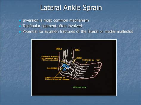
The present study aimed to evaluate the sensitivity and specificity of clinical tests and ultrasonography in detecting ankle ligament injuries. In this cross-sectional study, 105 patients with a history of ankle sprain were included. Ankle ligaments, including syndesmosis of ankle, as well as deltoid, calcaneofibular, anterior talofibular, and posterior talofibular .The three ligaments that make up the lateral collateral complex are the anterior talofibular ligament (ATFL), the calcaneofibular ligament (CFL) and the posterior talofibular (PTFL), and they are typically injured in this order during an inversion sprain.

Talofibular Ligament Injury Clinical Presentation
Intraoperative view of the lateral ankle ligaments (CFL and ATFL) along with the peroneal sheath and inferior extensor retinaculum (IER). Ankle stability is conferred by passive ligamentous restraints, articular surface congruity, and active musculotendinous units. 33 The ATFL, CFL, and PTFL provide static restraint to the lateral aspect of the ankle.
Radiopaedia.org
Fibular Ligament (PTFL): running posteriorly from the lateral malleolus to the posterior aspect of the talus; Calcaneo-Fibular ligament (CFL): running from the lateral malleolus to the lateral aspect of the calcaneous, in the middle between the ATFL and the PTFL . Squeeze Test or Thompson’s Test. To confirm a suspected Achilles tendon . The primary concerns are that existing clinical tests often fail to identify microinstabilities of the ankle joint complex; which consists of the anterior talofibular ligament (ATFL), calcaneofibular ligament (CFL), and the posterior talofibular ligament (PTFL). 22 Also, few tests target the primary stabilizers of the subtalar joint, consisting . Lateral ankle instability is a complex condition that can, at times, prove difficult to evaluate and treat for general practitioners. The difficulty in evaluation and treatment is due in part to the ankle complex is composed of three joints: talocrural, subtalar, and tibiofibular syndesmosis. All three joints function in conjunction to allow complex motions of the ankle joint.Imaging Tests. Imaging tests such as MRI (Magnetic Resonance Imaging) or ultrasound are used to diagnose ATFL injuries by providing detailed views of soft tissues and ligaments in the ankle. These tests help confirm the extent of ligament damage and help understand whether it's a bone-related injury or something related to the tissues and muscles.
Acute ankle sprains are commonly seen in both primary care and sports medicine practices as well as emergency departments and can result in significant short-term morbidity, recurrent injuries, and functional instability. Although nonoperative treatment is often successful in achieving satisfactory outcomes, correct diagnosis and treatment is important at the time of .
Posterior talofibular ligament
Anterior talofibular ligament tear is one of the common types of ankle injuries. . calcaneofibular ligament, and posterior talofibular ligament (PTFL). The anterior talofibular ligament (ATFL) forms the anterior compartment of the ligament complex. . as well as any changes in the end feel, is observed. During this test, the ankle is flexed .

222K Followers, 61 Following, 4,624 Posts - See Instagram photos and videos from Telma Goncalves dos Santos (@telmasantos544)
tests for ptfl tear|Posterior talofibular ligament