vein compression test|catheterized varicose veins : custom A point-of-care ultrasound consisting of a limited evaluation with compression from thigh to knee (extended compression ultrasound [ECUS]) is appropriate when CDUS is not available in a timely manner. ECUS is favored . Play AUD-20231210-WA0003 by Doc Equi on audio.com and discover something new. Listen tracks or upload your own audio files for free.
{plog:ftitle_list}
webFNaF 1 Doom LITE foi criado por um brasileiro em cima do Five Nights at Freddy's 1 Doom Mod criado por @Skornedemon em cima de Doom usando GZDoom, Este mod foi feito .
Iliac vein compression syndrome, also called May-Thurner syndrome, is caused by anatomic compression of the veins in the pelvis (iliac veins), which reduces blood return from the legs. The iliac veins carry blood from each leg .

zx10r compression test
A point-of-care ultrasound consisting of a limited evaluation with compression from thigh to knee (extended compression ultrasound [ECUS]) is appropriate when CDUS is not available in a timely manner. ECUS is favored . To diagnose varicose veins, a healthcare professional might use a test called a venous Doppler ultrasound of the leg. It's a painless test that uses sound waves to look at . Tests used to diagnose or rule out DVT include: D-dimer blood test. D dimer is a type of protein produced by blood clots. Almost all people with severe DVT have increased .

what is a varicose vein test
In this section, we will go over a step-by-step ultrasound approach to evaluate for deep vein thrombosis by visualizing the veins and using venous compression using a standard DVT Ultrasound Protocol. There is not enough evidence to determine if compression stockings are effective in the treatment of varicose veins in the absence of active or healed venous ulcers. Interventional treatments.
Diagnosis. Treatment. Prevention. When your veins have trouble sending blood from your limbs back to the heart, it’s known as venous insufficiency. In this condition, blood doesn’t flow back. Duplex ultrasound scanning is now the gold standard and first diagnostic test for assessing the lower limbs in patients with suspected varicose veins or CVD. 3,6,9 Duplex . One of the common treatment options for varicose veins is compression therapy (with compression stockings or bandages). If a patient has a significant degree of arterial disease they may not be suitable for .Iliac vein compression syndrome. How does May-Thurner syndrome affect my body? May-Thurner syndrome makes it harder for blood to flow back to your heart. Instead, it may pool in your legs and can develop into deep vein thrombosis (DVT). DVT occurs when a blood clot forms in your deep leg veins. DVT symptoms may include:
varicose veins ultrasound
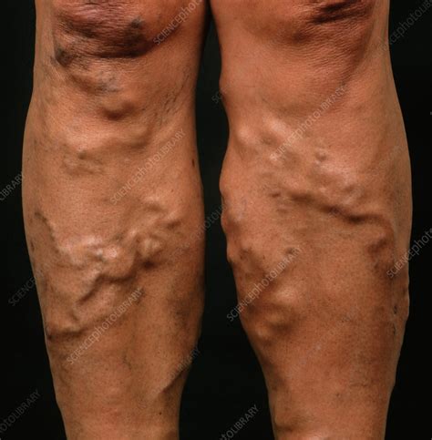
varicose veins in legs pictures
This noninvasive test uses sound waves to create pictures of how blood flows through the veins. It's the standard test for diagnosing DVT. For the test, a care provider gently moves a small hand-held device (transducer) on the skin over the body area being studied. . Support stockings (compression stockings). These special knee socks help .

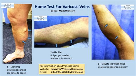
Varicose veins are subcutaneous veins dilated to at least 3 mm in diameter when measured with the patient in an upright position. They are part of a continuum of chronic venous disorders ranging .
Peripheral venous ultrasound (US) is commonly used to diagnose and evaluate venous disease, including acute thromboembolism, postthrombotic syndrome, and chronic venous insufficiency (see Image. Chronic Venous Insufficiency). Venous duplex ultrasound is quick to perform and easily accessible. It is noninvasive and offers no radiation risk to the .
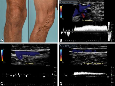
To diagnose varicose veins, a healthcare professional might use a test called a venous Doppler ultrasound of the leg. It's a painless test that uses sound waves to look at blood flow through the valves in the veins. . Self-care measures also might keep the veins from getting worse. Compression stockings. Wearing compression stockings all day .A study from 2011 showed that 75% of junior clinicians believed this vein was a part of the superficial venous system (Thiagarajah, Venkatanarasimha & Freeman). A clot in the (superficial) femoral vein is in fact a deep vein thrombosis and requires anticoagulant therapy to prevent a pulmonary embolus. Don’t get confused by the nomenclature! All ultrasound examinations were done using a uniform protocol that started with venous compression every 2 cm in the transverse plane of the common femoral. The great saphenous vein was examined at the saphenofemoral junction. . The two-point compression group included all patients with DVT locations detected by a two-point compression test.Varicose veins are swollen, bulging veins that often happen on the thighs or calves. . Wear compression stockings for a few days or weeks, if advised. These stockings gently squeeze your legs. This helps to prevent swelling in your legs. It can also help stop your blood from clotting or pooling. . Who will do the test or procedure and what .
Varicose veins are characterized by subcutaneous dilated, tortuous veins greater than or equal to three millimeters, involving the saphenous veins, saphenous tributaries, or non-saphenous superficial leg veins with age and family history considered important risk factors.[1] Varicose veins are considered a common clinical manifestation of chronic venous disease.[2] .Researchers don’t know what causes pelvic congestion syndrome. Still, problems with blood flow in your ovarian veins and the veins in your pelvis play a role. Normally, blood flows upward from your pelvic veins and toward your heart via the veins in your ovaries. Structures called valves in your veins prevent blood from flowing backward.
In this 1-minute video POCUS tip, learn how to perform a Venous Compression Test for DVT.Visit Us -- https://www.pocus.org/View Our POCUS Resources -- https:. Nutcracker syndrome is a type of extrinsic vein compression syndrome. In these syndromes, the structure of your blood vessels puts pressure on one of your veins. . They may not even know they have nutcracker phenomenon until their healthcare provider sees their anatomy on an imaging test. Nutcracker syndrome refers to the phenomenon when it .
Varicose veins are dilated superficial veins in the lower extremities. Usually, no cause is obvious. Varicose veins are typically asymptomatic but may cause a sense of fullness, pressure, and pain or hyperesthesia in the legs. Diagnosis is by physical examination. Treatment may include compression, wound care, sclerotherapy, and surgery. Iliac vein compression (LIVC) is a prevalent finding in the general population, but a smaller number of patients are symptomatic. ILVC should be considered in symptomatic patients with unexplained unilateral lower leg swelling. Patients typically complain of one or more of the following symptoms: lower leg pain, heaviness, venous claudication . Deep venous thrombosis (DVT) is a common condition that appears in the emergency department and outpatient settings. Clinical diagnosis is unreliable due to the infrequency of the classic findings of edema, warmth, erythema, pain, and tenderness, which are present only in 23% to 50% of patients.[1] When a patient presents with findings consistent .
Jugular vein compression is a serious condition that can cause debilitating pain, fatigue, and dizziness. There are various treatment options available, ranging from conservative care to invasive surgeries. It’s essential to choose well for .
ultrasound for venous thrombosis
varicose veins, swollen, discolored, distended, or twisted veins just beneath the skin’s surface; . The easiest way to treat venous reflux is by wearing compression stockings. These can help . If a healthcare provider suspects a patient has deep vein thrombosis (DVT), a condition defined by a blood clot forming in one of the deep veins, they will attempt to make a definitive diagnosis as quickly as possible. There is a potential for such a blood clot to loosen and travel to the lungs, which can cause a potentially life-threatening pulmonary embolism (PE).below the knee - to assess incompetence between the short saphenous vein and the popliteal vein. [3] Superficial veins of the leg normally empty into deep veins, however retrograde filling occurs when valves are incompetent, leading to varicose veins. The test is named for Friedrich Trendelenburg, who described it in 1891. [4] Compression of the left renal vein can occur primarily in two anatomic locations 10. anterior nutcracker syndrome (classical): occurs at the branching of the SMA off of the aorta. posterior nutcracker syndrome (rarer form): a retroaortic left renal vein is compressed between the aorta and vertebrae.
The first test is usually a duplex ultrasound. . The most common treatment for venous insufficiency is prescription compression stockings. These special elastic stockings apply pressure at the .
May-Thurner syndrome is the compression of the iliac vein against the lumbar spine by an overlying iliac artery, resulting in venous insufficiency, stenosis, and obstruction. While May-Thurner syndrome may be asymptomatic, typical symptoms include pain, swelling, and skin changes in the ipsilateral lower extremity. There are several variants of May-Thurner . It expands the vein. After compression (D), the vein does not collapse but has an oval shape indicating an acute DVT based on the noncompressible but deformable vein. DVT indicates deep venous thrombosis. . (CDUS) is the preferred venous ultrasound test for the diagnosis of acute DVT. CDUS is compression of the deep veins from the inguinal .The areas of vein compression are listed in Table 2. Figure 1. Compression of the left common iliac vein (CIV) by the right common iliac artery (CIA) over the fifth lumbar vertebra (A). The vein velocity ratio is 5.8. Narrowing of the CIV is apparent with mosaic color due to aliasing from the high velocity. No flow is seen in the left CIV .
This is the most common test for diagnosing a DVT because it is non-invasive and widely available. This test uses ultrasound waves to show blood flow and blood clots in your veins. . You may also have swelling because the DVT is blocking blood flow in your vein. You wear most compression stockings just below your knee. These stockings are . Sometimes varicose veins are removed with surgery. For superficial thrombophlebitis, your doctor might recommend applying heat to the painful area, elevating the affected leg, using an over-the-counter nonsteroidal anti-inflammatory drug (NSAID) and possibly wearing compression stockings.Noninvasive diagnosis of deep vein thrombosis (DVT) is based on ultrasound examination of the leg veins, usually restricted to only compression of the proximal veins (CUS). Patients with negative CUS findings require a second examination or a combination with other tests, which impairs clinical effi .
ultrasound for calf veins
webSonhar com marido bêbado. Sonhar com marido bêbado sugere que você não está dando bons exemplos para alguém para a qual é uma referência. O marido é alguém que confiamos e que deve ter uma atitude positiva .
vein compression test|catheterized varicose veins