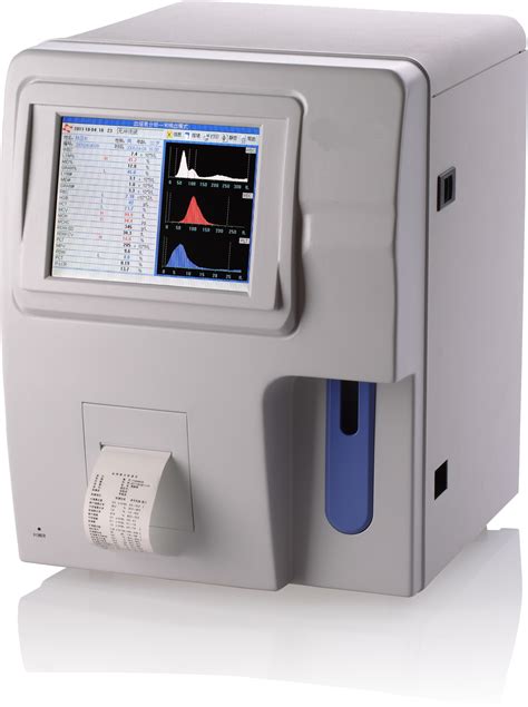
digital cell image analyzers in the hematology laboratory|Digital morphology analyzers in hematology: ICSH review and : distributor Digital image analyzers also allow remote networked laboratories to transfer images rapidly to a central laboratory for review, and facilitate a variety of essential work functions in laboratory . Resultado da 22 de fev. de 2024 · History the iron head of a tilting spear, used as a lance in jousting.. Click for English pronunciations, examples sentences, video.
{plog:ftitle_list}
Sou um vaso, sou de barro, e o Oleiro, me fez assim Sou de .
Digital morphology analyzers in hematology: ICSH review and
e18 standard test methods for rockwell hardness of metallic materials
Digital cell image analyzers in the hematology laboratory
The DM Systems (further referred as CellaVision) (CellaVision AB) are automated digital image analyzers that locate cells in a blood smear, capture images of the cells, .The digital morphology applied to hematology is based on the . Using a digital cell image analyzer, a laboratorian can view cells that have been pre-classified by the software to improve the accuracy of cell identification and proper .
Digital image analyzers also allow remote networked laboratories to transfer images rapidly to a central laboratory for review, and facilitate a variety of essential work functions in laboratory .Automated white blood cell differential counts and digital images of white blood cells. . Performance evaluation of the latest fully automated hematology analyzers in a large, commercial laboratory setting: a 4-way, side-by-side .Scopio X100 and X100HT meet your lab’s needs for a digital cell morphology analysis suite with Full-Field PBS imaging and AI-powered decision support. . White Blood Cells DSS for detection and pre-classification into 14 classes. .

1. Genc S, Dervisoglu E, Erdem S, Arslan O, Aktan M, Omer B. Comparison of performance and abnormal cell flagging of two automated hematology analyzers: Sysmex XN 3000 and Beckman Coulter DxH 800. Int J Lab Hematol. 2017 Dec;39(6):633-640. doi: 10.1111/ijlh.12717. Epub 2017 Oct 4. PMID: 28980399. Beckman Coulter is a distributor of Scopio Labs. The evolution of complete blood count (CBC) methodology from manual calculations to sophisticated high throughput hematology analyzers is the focus of this article. In recent years, hematology testing has greatly benefitted from the combination of various technologies with automated neural networks. In addition to an increasing complexity of the .
e450 hard drive fails self tests
The MC-80 is taking digital morphology analysis to the next level, delivering clearer images which are able to capture abnormalities in more detail. With advanced algorithms, the analyzer enables better identification of different cells with high throughput, resulting in greater productivity. Alferez S, Merino A, Mujica LE, Ruiz M, Bigorra L, Rodellar J. Automatic classification of atypical lymphoid B cells using digital blood image processing. Int J Lab Hematol. 2014;36(4):472-480. Gao Y, Mansoor A, Wood B, Nelson H, Higa D, Naugler C. Platelet count estimation using the CellaVision DM96 system. J Pathol Inform. 2013;4:16-3539.114207. We read the article “Digital morphology analyzers in hematology: ICSH review and recommendations” by Kratz et al1 published in this journal with great interest. While publication of a review article and recommendations for such a new and exciting technology was timely, there seems to have been some misunderstandings in the article which we would like . Background and Significance. Cell morphology is a cornerstone of hematological diagnosis. 1 Interpretation of peripheral blood smear (PBS) is a complex process involving cell recognition and classification, identification of abnormal cellular elements, weighing of those abnormalities relative to each finding and the overall interpretation of the finding, ultimately .

Bigorra L, Merino A, Alférez S, Rodellar J. Feature analysis and automatic identification of leukemic lineage blast cells and reactive lymphoid cells from peripheral blood cell images. J Clin Lab .Introducing a revolution in digital cell morphology that will transform the way you see, work and diagnose. . or zoom in on the smallest details all at an unprecedented resolution of 100X. With Scopio’s full-field imaging, lab teams, hematopathologists and clinicians can now get the complete picture, vital for confident clinical decision . In laboratory hematology, static, single-frame digital images are widely used in education, automated cell image analyzers, and for research applications. Recently, scanning microscopes that can capture virtual slides at high resolution have enabled the creation of high quality hematology virtual slides.
Rapid and accurate counts of red blood cells (RBCs), nucleated RBCs, platelets, and white blood cells (WBCs) (total and differential WBCs) are important requirements for a hematology laboratory. . Digital image analysis of blood cells Clin Lab Med. 2015 Mar;35(1) :105 . Automated blood cells analyzers; CellaVision; Differentials; Digital .
Background The Sysmex DI-60 system (DI-60, Sysmex, Kobe, Japan) is a new automated digital cell imaging analyzer. We explored the performance of DI-60 in comparison with Sysmex XN analyzer (XN .
Europe PMC is an archive of life sciences journal literature. We read the article “Digital morphology analyzers in hematology: ICSH review and recommendations” by Kratz et al1 published in this journal with great interest. While publication of a review article and recommendations for such a new and exciting technology was timely, there seems to have .
This study aims to evaluate the white blood cell differential performance of the Mindray MC‐80, the new automated digital cell morphology analyzer. The manual differential count has been recognized for its disadvantages, including large interobserver variability and labor intensiveness. In this light, automated digital cell morphology analyzers have been .
Introduction. Since the previously published ICSH guidelines in 1994, haematology analysers have evolved greatly. Automated analysers speed up the workflow in the laboratory and improve precision as more cells are counted and cell classifications are based on more measured objective properties (light scatter, fluorescence, digital imagine, etc.). In addition, state-of-the-art data for digital morphology analyzers are still unavailable in the literature. In particular, Vis and Huismans 7 offer state-of-the-art precision values for hematology analyzers that count thousands of cells, and maybe this criterion is not suitable for digital image analyzers, which count only 200 cells. 1. Introduction. Morphological assessment of peripheral blood smears is crucial for disease screening and diagnosis in clinical laboratories. When blood samples are flagged with abnormalities by automated hematology analyzers according to defined verification rules, or when there are clinical concerns [1], [2], a blood smear examination is typically performed as . The Scopio Labs Full-Field X100 PBS system with remote analysis capacity significantly reduced PBS TAT and improved the morphology workflow of the hematology laboratory. Abstract Background The demand for morphological diagnosis by peripheral blood smear (PBS) analysis with clearly defined turnaround times (TAT), coupled with a shortage of .
The system leverages proven digital image analysis technology to locate and examine cells in blood and other body fluids, saving time, accelerating turnaround, and increasing technologists' productivity throughout high-volume labs. In addition, the CellaVision DM9600 system easily adapts to any hematology workflow within hospital IT environments.
are automated digital image analyzers that locate cells in a blood smear, capture images of the cells, preclassify the cells using image analysis software, and then display the images on a computer screen. The analyzer scans a portion of a microscopy slide and automatically identifies an appropriate analysis area (monolayer) in which it locatesare automated digital image analyzers that locate cells in a blood smear, capture images of the cells, preclassify the cells using image analysis software, and then display the images on a computer screen. The analyzer scans a portion of a microscopy slide and automatically identifies an appropriate analysis area (monolayer) in which it locates Background: Digital morphology (DM) analyzers are increasingly being used for white blood cell (WBC) differentials. We assessed the laboratory efficiency of the Sysmex DI-60 system (DI-60; Sysmex .
Simplifying Digital Cell Morphology for your lab. CellaVision DC-1 is a revolutionary hematology analyzer that is optimized to simplify blood cell differentials in diagnostic labs. It effectively automates and simplifies the work that is traditionally done by manually using conventional microscopy. . The ability to archive images enables . In laboratory hematology, static, single-frame digital images are widely used in education, automated cell image analyzers, and for research applications. Recently, scanning microscopes that can capture virtual slides at high resolution have enabled the creation of high quality hematology virtual slides.
WEBApós o último ataque em Woodsboro, Sam, Tara e os outros sobreviventes partem para Nova York. Tentando seguir com suas vidas, eles se tornam alvo de um novo Ghostface. Assista online Pânico VI pelo Globoplay. Acesse agora mesmo!
digital cell image analyzers in the hematology laboratory|Digital morphology analyzers in hematology: ICSH review and