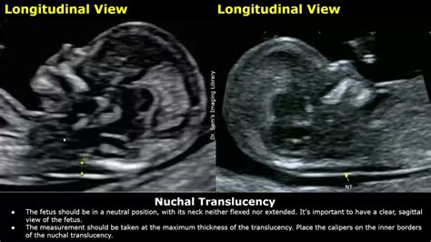
how to measure nuchal fold thickness|nuchal fold measurement chart : wholesale Second trimester thickened nuchal fold has a high specificity for aneuploidy. ACOG and SMFM define an abnormal nuchal fold as ≥ 6mm between 15 and 20 weeks of gestation. It is the most powerful second . WEBAt a time when standard setters are expanding the overall definition of who should be treated as “politically exposed,” for example by including domestic as well as foreign PEPs, it is important that FIs still focus the greatest effort on those PEPs who pose the very highest corruption risk, i.e. those genuinely in senior or prominent public positions and .
{plog:ftitle_list}
JANE IS OPEN ALL WEEK LONG FOR BRUNCH AND DINNER. ×. 100 West Houston St, New York, NY 10012 (212) 254-7000. Hours & Location. Menus. Private Dining. Promotions. Delivery.
Measuring nuchal skin fold thickness after tilting the transducer towards the occiput approximately 30° while maintaining the appropriate intracranial landmarks. The . Second trimester thickened nuchal fold has a high specificity for aneuploidy. ACOG and SMFM define an abnormal nuchal fold as ≥ 6mm between 15 and 20 weeks of gestation. It is the most powerful second .The nuchal fold is a normal fold of skin at the back of a baby’s neck. This can be measured between 15 to 20 weeks in pregnancy as part of a routine prenatal ultrasound. The nuchal fold .Objective To assess the effect of imaging angle and fetal presentation on the measurement of nuchal skin fold thickness (NFT) in the second trimester. Methods Fetal NFT was .
ABSTRACT. Objective To assess the effect of imaging angle and fetal presentation on the measurement of nuchal skin fold thickness (NFT) in the second trimester. Methods Fetal .The nuchal translucency test measures the nuchal fold thickness. This is an area of tissue at the back of an unborn baby's neck. Measuring this thickness helps assess the risk for Down .
A nuchal translucency (NT), also called nuchal fold or nuchal thickness, is a measurable area at the back of the fetal neck. It is examined using ultrasound as part of combined screening for .The observed values of nuchal fold thickness and biparietal diameter can easily be used as coordinates to identify the corresponding likelihood ratio. The likelihood ratio can then be multiplied by the prior probability according to .Nuchal fold can be spuriously thickened by angling caudally (intersecting the inferior level of the cerebellum and occiput). This nuchal skin fold increases with advancing gestational age .Ultrasound is the go-to method for measuring nuchal fold thickness. It’s non-invasive and gives healthcare professionals a clear view of your little one’s development. . Is nuchal fold measurement at 20 weeks different for twins .
Nuchal fold measurement is obtained from the outer edge of the occipital bone to the skin surface in the transaxial plane of the fetal head at the level of the cavum septi pellucidum and cerebellar hemisphere. . , Fassnacht MA et.al. Routine measurement of nuchal thickness in the second trimester. J Matern Fetal Med 1992; 1:82-86 ;
when to measure nuchal fold
The nuchal translucency test measures the nuchal fold thickness. This is an area of tissue at the back of an unborn baby's neck. Measuring this thickness helps assess the risk for Down syndrome and other . Your health care provider uses abdominal ultrasound or a vaginal ultrasound to measure the nuchal fold. All unborn babies have some fluid .What is Nuchal Fold Thickness. The Nuchal fold Thickness is a regular fold of skin found at the back of the fetal neck during the second trimester of pregnancy. Increased nuchal fold thickness is a soft indicator associated with a variety of fetal abnormalities and is measured during a regular second-trimester ultrasound.The Nuchal Fold Calculator uses a specific formula to determine the thickness of the nuchal translucency (NT) or nuchal fold, which is a space at the back of the fetal neck. The formula involves measuring the NT thickness during an ultrasound examination, typically performed between the 11th and 14th weeks of pregnancy. The formula is as follows:A nuchal translucency (NT) scan is an ultrasound scan to measure the amount of fluid at the back of your baby's neck. It is part of the combined screening test for Down's syndrome, which is offered at around 12 weeks of pregnancy. The results of your NT scan are combined with blood test results and other factors, such as your age.
Nuchal translucency (NT) is a measure of a thickness of a fold located on the fetuses' neck. This fold's greater thickness is connected to the greater prevalence of genetic disorders, fetal death, and its major abnormalities.NT is one of the 1st trimester screening methods.. We perform a nuchal translucency test during the ultrasound examination in the .
An abnormal measurement is when the skin fold measure is larger than the normal range of up to 2 mm at 11 weeks or 2.8 mm at 13 weeks 6 days. Your doctor will consider the measurements along with .
The nuchal translucency test measures the nuchal fold thickness. This is an area of tissue at the back of an unborn baby's neck. . Your health care provider uses abdominal ultrasound or a vaginal ultrasound to measure the nuchal fold. All unborn babies have some fluid at the back of their neck. In a baby with Down syndrome or other genetic .
thick nuchal fold normal baby
nuchal translucency normal range at 13 weeks
nuchal thickness chart
What is Nuchal Translucency (NT)? NT is the name given to the black area seen by ultrasound at the back of the fetal head/neck between 11 - 14 weeks of gestation. . There is a screening test, called the combined test, that determines this risk calculation. The test combines the measurement of the NT, the length of the baby, your age, and the .The thickness of the nuchal fold is expected to fall under a certain level during your first trimester Nuchal Translucency scan, and the abnormal measurement of the same would indicate a genetic disorder. What Causes Abnormal Nuchal Fold Thickness? The increased nuchal fold thickness might be the cause of lymphatic obstruction.
To assess the association of adverse pregnancy outcomes in fetuses with increased nuchal folds in the setting of normal genetic testing. Increased measurement of the nuchal fold (≥ 6 mm from 14 weeks to 22 weeks of gestational age) is considered a soft marker for chromosomal aneuplodies, as well as for structural defects in the fetus, most commonly cardiac defects. We .
I just got back from my 20 week scan. It looks like baby is measuring big, but everything looked normal except for a nuchal fold measurement of 7mm. We originally opted out of genetic testing but after that measurement decided to go forward.
The measurement of the nuchal fold is a test where the accumulation of liquid in the neck of the fetus is evaluated by ultrasound. . Normally, this thickness is less than 3 mm. When the nuchal translucency is above this value, it can be associated with an increased risk of certain genetic diseases, such as Down syndrome.First, nuchal fold thickness measurement (typically performed between 16-20 weeks) must be differentiated from the nuchal translucency measurement done in the first trimester. Unlike other "soft markers" for chromosome abnormalities .The requirements for obtaining the FMF certificate of competence in the nuchal translucency (NT) scan are: Attendance of the internet based course on the 11-13 weeks scan. Submission of a logbook of 3 images demonstrating the measurement of NT. Protocol for measurement. The gestational period must be 11 to 13 weeks and six days.
Objective: To establish normal values of fetal nuchal fold thick-ness at 14-16 weeks of gestation by transvaginal sonography. Methods: Transvaginal sonography was used to measure nuchal fold thickness in 182 normal pregnancies at 14-16 weeks of gestation. Nuchal fold thickness was measured as the distance from the outer skull bone to the outer skin surface in the .The nuchal translucency (NT) is an ultrasound measurement defined as the collection of fluid under the skin behind the neck of the fetus obtained between 10 and 14 weeks’ gestation (crown–rump length between 38–45 and 84 mm) (Fig. 12.1).While some fluid is present in the nuchal space of all fetuses, regardless of chromosomal status, it tends to increase among .Prenatal ultrasound: Increased nuchal fold (2nd trimester)
The requirements for obtaining the FMF certificate of competence in the nuchal translucency (NT) scan are: Attendance of the internet based course on the 11-13 weeks scan. Submission of a logbook of 3 images demonstrating the measurement of NT. Protocol for measurement. The gestational period must be 11 to 13 weeks and six days.
The nuchal fold normally measures less than 6mm at 20 weeks. What does this mean? It can be a normal variation or associated with extra fluid in the skin (oedema) which may be due to an infection or a chromosomal condition such as Downs’s syndrome (Trisomy 21). .The nuchal translucency test is an ultrasound analysis that consists of measuring the nuchal fold of the fetus. . In addition, it is said that the risk increases as the thickness of the nuchal translucency increases, making it known that there is a risk of 10% if the thickness is between 3-4 mm, the risk is 40% if the measure is located .
custom garden soil moisture humidity and ph acidity meter
Definition The nuchal fold thickness is a measurement taken during a fetal ultrasound. It refers to the amount of skin observed at the back of a fetus’s neck, ideally between the 18th and 20th weeks of pregnancy. Increased thickness can be an indication of certain genetic disorders, like Down syndrome. Key Takeaways The term ‘nuchal [.]
Nuchal Fold Calculator Basic Calculator Advanced Calculator Crown-Rump Length (mm) Nuchal Fold Thickness (mm) Crown-Rump Length (mm) Nuchal Fold Thickness . This measurement can help doctors assess the risk of certain genetic disorders, such as Down syndrome, as a thicker nuchal fold can be a potential indicator of these conditions. However .Objective: To assess the effect of imaging angle and fetal presentation on the measurement of nuchal skin fold thickness (NFT) in the second trimester. Methods: Fetal NFT was prospectively measured in 921 women at 18-21 weeks' gestation. The population was divided into two groups according to fetal presentation. Group A comprised 643 fetuses in cephalic or transverse .They'll then measure the width of the nuchal fluid at the back of your baby’s neck. The skin will appear as a white line, and the fluid under the skin will look black. You'll usually be able to see your baby's head, spine, limbs, hands and feet on the screen. Your sonographer will be able to rule out some major abnormalities, such as problems . Accumulated local effect (ALE) plot of the top four features—(a) Nuchal Fold, (b) Uterine RI, (c) Uterine PI, and (d) Abdominal Circumference from the Support Vector Machine (SVM) in predicting FGR. Nuchal fold below 3.7 mm, Ut RI above 0.5, Ut PI above 1.59, and AC below 158 mm showed an increased probability of fetuses being born as FGR.

custom garden soil moisture meter lowes
WEB15 de jul. de 2021 · Como Tocar Segredos no Violão, Frejat (Aula de Violão Simplificada) Seja Meu Aluno no Meu Curso Online de Violão: https://tocandofacilonline.com/yt ️Apostila.
how to measure nuchal fold thickness|nuchal fold measurement chart