how to measure gallbladder wall thickness on ultrasound|gallbladder wall thickness 2mm : online sales Thick gallbladder wall is a common finding seen on ultrasounds done of the gallbladder. There are many causes of a thick gallbladder wall that range from benign all the .
Resultado da Massagens no Porto eróticas e relaxantes. Disfrute do prazer de receber uma massagem de uma mulher sensual. Anuncios de massagistas em Portugal.
{plog:ftitle_list}
Resultado da Milfy City V1.0 Final Edition - Preview 9/15. lolz guy's been saying you'll get the finished version for over a year now. he's just trying to see how long he .
normal gallbladder wall thickness ultrasound
Ultrasound depicts diffuse wall thickening of a stone-free painless gallbladder and large-caliber hepatic veins (arrowheads) and inferior vena cava, as supporting evidence of . Normal findings on a gallbladder ultrasound include a thin-walled (<3 mm), anechoic, and pear-shaped structure that typically measures between 7–10 cm in length and . Ultrasound is the primary imaging technique used to assess gallbladder wall thickness. It’s a safe, non-invasive, and effective method for visualizing gallstones, polyps, and .
• anterior GB wall thickness (measured in the short axis) o f more than 3 mm, • and pericholecystic fluid (free fluid around the gallbladder). How to do it better: • Placing the patient .
Diffuse Pattern. Diffuse gallbladder wall thickening (> 3 mm by ultrasound) can be seen in such primary gallbladder inflammatory processes as acute, chronic, and acalculous .
Thick gallbladder wall is a common finding seen on ultrasounds done of the gallbladder. There are many causes of a thick gallbladder wall that range from benign all the . Another measurement needed for a complete biliary POCUS is a measurement of the gallbladder wall. The normal gallbladder wall appears as a pencil-thin line, with 3 to 4 mm being the upper limit of normal thickness for .Gallbladder (GB) wall normally appears as a pencil-thin line [1, 2]. Most studies suggest a wall thickness of 3 mm as the upper limit of normal thickening [3, 4]. In a retrospective review of 4119 patients, it was found that the GB wall in .
Ultrasound. As the gallbladder was low-hanging fruit for the first, rudimentary B-mode ultrasound scanners, normal limits of the gallbladder wall thickness (3 mm) were established early on 1. . Most commonly the .
A thick gallbladder wall is defined as one that is greater then 3 mm in thickness. The gallbladder has to be distended in order to correctly measure the wall thickness. A contracted gallbladder after a meal may have a thick wall simply from being contracted. Although less sensitive than ultrasound, CT may also show gallbladder wall thickening or distention, cholelithiasis, . The thickness of the gallbladder wall depends on the degree of gallbladder distention, and pseudothickening can occur in the postprandial state. . malignant lesions are usually smaller than 1 cm. Polyps measuring 5–10 mm .
Gallbladder wall thickness; Pericholecystic free fluid; Common Bile Duct (CBD) measurement Measure from inner to inner; Sonographic Murphy's sign Pain with ultrasound probe palpation over gallbladder; Normal Findings. No gallstones; Gallbladder anterior wall <4 mm; No pericholecystic free fluid; Common bile duct < 4 mm plus 1mm per decade after . Gallbladder wall thickening can be measured in the short or long axis and should be measured at the thickest point along the wall; A number of normal states can also cause gallbladder wall thickening – including congestive states, ascites, and in the setting of a contracted gallbladder. Ultrasound findings must be interpreted in conjunction . Results: The relationship between the gall-bladder wall thickness and the occurrence of intraoperative difficulties in the analysed set is deterministic (AUC = 1), and the wall thickness of ≥ 5 mm allows to predict their occurrence as soon as at the stage of diagnostic evaluation (p < 0.001). In addition, the ultrasound-measured GB wall . Objectives To evaluate the association between ultrasound assessment of gallbladder wall thickness (GBWT) among severe dengue patients and dengue patients with warning signs to their clinical outcomes. Methods A prospective, cross-sectional study involving adult dengue patients presented to our emergency department between March until .
Ultrasonographic Measurements of the Liver, Gallbladder Wall Thickness, Inferior Vena Cava, Portal Vein and Pancreas in an Urban Region, Malaysia. . The aim of this study is to determine the differences of ultrasound measurement of the liver length, gallbladder wall thickness (GBWT), IVC, PV, and pancreas among genders and between normal and .Measurement of gallbladder lesions. The examples in section 1 indicate that a reliable measurement of the thickness of the gallbladder wall is only possible in the hepatic wall. It is advisable to measure it on an enlarged sonogram using the zoom function (Fig. 12). Fig. 12.

normal gallbladder wall thickness
The purpose of this work was to evaluate, by ultrasonographic method, gallbladder wall thickness and gallbladder emptying after a standard meal in normal subjects and in patients with compensated liver cirrhosis without gallstones. Methods. Twenty-three patients with Child-Pugh class A liver cirrhosis and twenty healthy controls were studied. A prospective ultrasound study of gallbladder wall thickness in patients with suspected gallbladder disease was performed over a 9 month period. Gallbladder walls thicker than 3 mm in fasting . As the thin hyperechoic serosa is rarely discernible, bowel wall thickness is measured from the outer contour of the ventral muscularis to the outer contour of the dorsal muscularis, and then of course, divided by two. Normally, single small bowel wall thickness during compression is about 1.5 - 2.5 mm. The Gallbladder Reporting and Data System (GB-RADS) ultrasound (US) risk stratification is proposed to improve consistency in US interpretations, reporting, and assessment of risk of malignancy in gallbladder wall thickening in non-acute setting. It was developed based on a systematic review of the literature and the consensus of an international multidisciplinary .
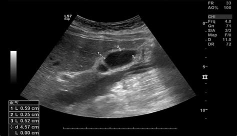
Figure 2. Anatomy of the gallbladder. Impression: Normal gallbladder . How to Measure Gallbladder Wall Thickness using B-mode ultrasound. Measure the wall of the gallbladder as shown. Measure the wall of the gallbladder closest to the anterior abdominal wall in the region of the liver anteriorly and the bile filled lumen of the gallbladder .
Diffuse gallbladder wall thickening is commonly encountered in diagnostic settings. The ability of ultrasonography, computed tomography (CT), and magnetic resonance imaging (MRI) to directly visualize the thickened . Ultrasound for the Gallbladder . How to do it: 1. Patient is in the supine position, or left lateral decubitus position. . Edge artifact can make the GB wall appear thickened, thus only measure the GB wall thickness anteriorly in the short axis (where it looks round). d) Sludge in the GB can be mistaken for stones, but more commonly, artifact .A greater degree of gallbladder wall thickness is associated with an increased risk of conversion, increased postoperative complications, and longer lengths of stay. Classifying patients according to degree of gallbladder wall thickness gives more accurate assessment of the risk of surgery, as well . Background: Diffuse gallbladder (GB) wall thickening is caused by elevated systemic venous pressure, such as heart failure (HF). This study investigated the relationship between GB wall thickness (WT) and HF, and the prognostic impact of GBWT. Methods and Results: This prospective study included 116 patients with HF and 11 healthy controls. Among .
Each patient's preoperative evaluation included a gallbladder ultrasound, which included measurement of the diameter of the gallbladder wall. The entire series of cholecystectomies was evaluated according to the ultrasound measured diameter of the gallbladder wall. A thin gallbladder wall was less than 3 mm in diameter. The normal gallbladder wall appears as a pencil-thin line, with 3 to 4 mm being the upper limit of normal thickness for the anterior gallbladder wall. The anterior wall, closest to the transducer footprint, is measured due to an artifact called posterior acoustic enhancement leading to an artificially thick appearance of the posterior .
Differences of mean liver length, gallbladder wall thickness, inferior vena cava, portal vein, and pancreas between males and females. The differences of mean liver length, GBWT, IVC, PV, and pancreas between males and females were tested and illustrated in Table 2.The liver length was larger in males (12.2 ± 1.5 cm) than in females (11.7 ± 1.5 cm).To date, gallbladder wall thickness typically has been evaluated against an anecdotally reported measurement, without a formal evidence base. 4 Gallbladder wall thickness identified in our study is considerably less than the widely cited 3 mm value, 4 and more aligned with recent indirect evidence presented above 7, 8, 10 and the statement that . In the results of this study, the gallbladder wall thickness in TWGC patients was over 9.5 mm on average, which was significantly thicker than that in patients with chronic cholecystitis. e normal .

Ultrasound is commonly used to measure gallbladder wall thickness in dogs, with several reference values reported at 1–2 mm (Kealy et al. 2011) . Ultrasound of gallbladder wall thickening and its relation to cholecystitis. AJR Am J Roentgenol. 132(4):581–585. doi: 10.2214/ajr.132.4.581. .
The thickening of the gallbladder wall in patients with ascites is commonly related to hypoalbuminemia and/or portal hypertension. To evaluate the pathogenetic role of these two factors, we correlated gallbladder wall thickness (GBWT) with the albuminemia and the serum-ascites albumin gradient (SAAG), an index of portal hypertension, in 47 patients with ascites .
Although less sensitive than ultrasound, CT may also show gallbladder wall thickening or distention, cholelithiasis, . The thickness of the gallbladder wall depends on the degree of gallbladder distention, and pseudothickening can occur in the postprandial state. . malignant lesions are usually smaller than 1 cm. Polyps measuring 5–10 mm . Measure inner wall to inner wall. What to Look For [3] Gallstones -- especially in the neck as this is where they're most problematic . Sonographic Murphy's sign (maximal pain elicited when probe is pressing directly on the gallbladder) **One of the most important sonographic signs of cholecystitis [4] Wall thickening > 3mm (measure anterior wall)
Resultado da The base game is fairly straightforward, unfolding across a 5x3 grid with 20 active paylines on each spin. You can play the slot for as little as $0.20 .
how to measure gallbladder wall thickness on ultrasound|gallbladder wall thickness 2mm