how to measure placental thickness|placental thickness normal range : trader Sonographic criteria for detection of placenta accreta include placental lacunae, thinning of the retroplacental myometrium, and irregularity or disruption of the bladder-serosal interface, with bridging vessels between the . HellBoy. The Hellboy slot machine is developed based on the comic book of the same name. The creator of the game is the company Microgaming. Gamblers are given the opportunity to spin 5 reels with 20 adjustable paylines, winning sums with the coefficients of up to 10,000. In the slot, there is a wild symbol, a scatter, and a risk game.
{plog:ftitle_list}
Resultado da 21 de jul. de 2021 · Encontra aqui os melhores casinos online legais de 2024, onde podes jogar os melhores clássicos .
salt spray test cabinet
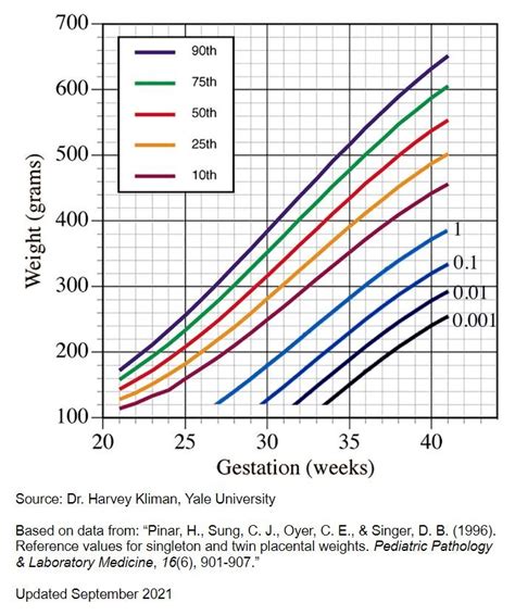
placental weight by gestational age
The maximum thickness of a normal placenta at any point during pregnancy is often considered to be 4 cm. Anterior placentas are ~0.7 cm thinner than posterior placentas and maximum thickness for an anterior placenta is ~3.3 cm 7. Ultrasound (USG) remains the preferred method for detecting placental abnormalities due to its benefits. This study aimed to evaluate placental thickness by USG in .EPV can be measured from 7 to 40 weeks. Use widest angle probe for large placentas. Beyond 36 weeks the placenta may be too large to visualize the entire width. Ideal measurements: .
placental thickness normal range
Sonographic criteria for detection of placenta accreta include placental lacunae, thinning of the retroplacental myometrium, and irregularity or disruption of the bladder-serosal interface, with bridging vessels between the .
Measuring the placenta is quick, easy, inexpensive and noninvasive with EPV. Three simple measurements taken during a standard prenatal ultrasound produce an Estimated Placental . We sought to determine the normal sonographically measured placental thickness in millimeters at the second-trimester scan (18 weeks to 22 weeks 6 days) and determine .
During the examination, the size, shape, consistency and completeness of the placenta should be determined, and the presence of accessory lobes, placental infarcts, hemorrhage, tumors and.Ultrasound (USG) remains the preferred method for detecting placental abnormalities due to its benefits. This study aimed to evaluate placental thickness by USG in various GA subgroups .
Placental thickness was measured at 24 and 36 weeks by ultrasound and was divided into three groups: Group A (normal placenta), Group B (thin placenta), and Group C (thick placenta); . The placental thickness (PT) can be used to estimate gestational age (GA). GA is frequently over or underestimated, as the conventional gestational estimation is based on . Placental thickness in the second trimester: a pilot study to determine the normal range. J Ultrasound Med 2012;31(2):213–218. Medline Google Scholar. . A new method for measurement of placental elasticity: .Regular ultrasound is used to evaluate placental condition – in particular “CTUP” (combined thickness of uterus and placenta also sometimes referred to as “CUPT” – combined uterine and placental thickness), the degree of .
Measurement of the combined thickness of the uterus and placenta (CTUP) and evaluation of the relationship between the chorioallantoic membrane and uterus of the mare are used to screen for placentitis and placental separation. The study was done to measure the placental thickness (PT) in pregnant women and find its correlation with the gestational age (GA) of the fetus by ultrasonography. Comparisons were also made with the other fetal biometry parameters, and baseline data were generated with respect to the gestational weeks and placental position.measure, Placental thickness of 2.5 cm to 3.75 cm is taken as normal. A placental thickness of ≥ 4 cm is regarded as abnormal [1]. It is documented that placental weight in a normal pregnancy at term is about one-fifth of the fetal weight. placental thickness is the simplest placenta percreta (the bladder should be mildly distended if evaluating for percreta on MRI) Other situations in which it may be useful. abnormal placental location; delineating bilobed placenta or accessory placenta; placental vascular anomalies; Practical points. mild placental lobulation and myometrial thinning can be seen in normal .
Endometrial thickness is a commonly measured parameter on routine gynecological ultrasound and MRI. The appearance, as well as the thickness of the endometrium, will depend on whether the patient is of reproductive age or postmenopausal and, if of reproductive age, at what point in the menstrual cycle they are examined. . Measurement .Why is placenta size important? Just as research has shown that SGA (small for gestational age) or IUGR (intrauterine growth restriction) fetuses are at increased risk for poor outcomes, including low APGAR scores at birth, NICU admission or stillbirth, a small placenta is also a risk factor that has been linked to a greater likelihood of poor outcomes.Normal placental thickness rarely exceeds 4 cm (1). Beware of fundal or lateral placental location in order to avoid measuring placental thickness in a tangential section. Avoid including a myometrial contraction in the measurement. Amount of amniotic fluid bears a relationship to apparent placental thickness. placenta, with linear echoes emerging at right angles. Because a tangential measurement would distort the placental thickness, the transducer was positioned on the abdomen perpendicular to the chorionic and basal plates after locating the cord insertion site and placental myometrial interface. The placental thickness was
variability in placental thickness was between patients. Unadjusted, the overall mean placental thickness was 24.6 (SD, 7.29) mm. The raw data stratified by both the week of gestation and the position of the placenta are shown in Table 2. One patient had a placental thickness of 54.9 mm whose gestational age was 22.6 weeks, age 34.9
The study was done to measure the placental thickness (PT) in pregnant women and find its correlation with the gestational age (GA) of the fetus by ultrasonography. Comparisons were also made with the other fetal biometry parameters, and baseline data were generated with respect to the gestational weeks and placental position. .was carried out to measure placental thickness [two-dimensional ultrasound; Medison ultrasound with transabdominal 3.5 MHz probe]. The placental site had been determined in a longitudinal section using two-dimensional real-time mode. The thickness of the placental had been measured at the level of .
Objectives. We sought to determine the normal sonographically measured placental thickness in millimeters at the second-trimester scan (18 weeks to 22 weeks 6 days) and determine whether the measurement should be . This may be related to the short insertion site included in PT measurement in that study, as Hoddick et al. suggested that short placental insertion sites often spuriously increase the thickness of a normal placenta. In this study, measurement of PT was carried out perpendicular to the uterine wall and through the placenta at the site of .Fetal and placental weight ratios. High fetal weight compared to placental weight is another indicator that a baby may have been stillborn due to outgrowing his/her small placenta. A “normal” ratio is 6:1. As a general rule of thumb, an 8:1 . Electronic calipers were used to take the measurement of the placenta thickness at the insertion of the umbilical cord, on a plane perpendicular to the placental surface, from the chorionic plate to the beginning of the basilar-myometrial surface. A straight line .
Measurement of placental diameter & thickness. A mobile ultrasound machine was used to acquire data; SONOSITE M-Turbo (made in USA). Curvilinear probe frequency of 3.5 – 5mHz with the participant lying in supine position on the examination couch, coupling gel was applied on the abdomen after exposing it. Placental thickness & diameter were .The placenta was demonstrated to increase in thickness with advancing menstrual age. At no stage of pregnancy was the normal placenta greater than 4 cm in thickness. Potential pitfalls in measuring placental thickness are addressed, as well as potential causes of aberrations in placental thickness.
placental thickness measurements range
Background: The purposes of this study are to sonographically measure the placental thickness (PT) in normal fetuses; to correlate it with gestational age (GA), fetal growth parameters, and .
placental thickness and gestational age
The criteria for a "thickened placenta" vary between studies based on gestational age, placental location, measurement technique, and maternal or fetal factors. Whereas most suggest thickness exceeding 4 cm is pathologic, a review had a threshold of 6 cm in the third trimester to classify placentomegaly. Several maternal and fetal conditions . Despite these studies, the clinical importance of abnormally large or small placentas remains unclear. Placental thickness is the easiest placental measurement to measure and could therefore play a potential role in screening for complications during second‐trimester sonography. First, though, we need to establish what is normal. Introduction This study aimed to identify if placental thickness measured from MRI images correlated with placenta percreta in patients with placenta previa. Methods Placental thickness was retrospectively measured in 161 patients from July 2018 to August 2020. The measurements were performed at the thickest part of the placenta in the lower .
ness of the utero-placental unit of 1.26 " 0.33 cm has been reported in mares with normal term preg-nancies. Measurements should be taken avoiding areas of compression of utero-placental thickness by the fetus, using the ventral uterine vasculature as landmark. The utero-placental thickness is af-fected by numerous factors, which may reduce the
salt spray test chamber
The location of the placenta should be described as anterior or posterior. The presence of placenta previa should be excluded by measuring the distance from the caudal edge of the placenta to the inner cervical os. A measurement of less than 3 cm is concerning for placenta previa and should be further evaluated.Placentomegaly | Radiology Reference Article | Radiopaedia.orgEstimated Placental Volume (EPV) is a simple measurement that can be done via ultrasound while a baby is in utero to estimate the size of a placenta. It requires 3 measurements (width, height, and thickness) that can be entered into a free app called Merwin’s Calculator to determine the volume (cc) of a placenta.
small salt spray test chamber

Resultado da Estúdio de Tatuagem The Old Tattoo Art - Fica localizado na Rua Marselhesa 535, Bairro Vila Mariana - São Paulo - SP / O dono e Tatuador Giullianno .
how to measure placental thickness|placental thickness normal range