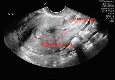
how to measure scar thickness on ultrasound|cesarean section scars chart : wholesaling This video talks about how to assess cesarean section scar by Ultrasound. WEB26 de nov. de 2020 · Md: Kate Kuray #KateKuray #KateKuray@female_model Поблагодарить можно через VK Pay или здесь: https://vk.com/app6471849_-107647545
{plog:ftitle_list}
Open World - STEAMUNLOCKED » Free Steam Games Pre .
LUS thickness measured by ultrasound during the third trimester of pregnancy is inversely correlated with uterine scar rupture/dehiscence at delivery. To better visualize the . This video talks about how to assess cesarean section scar by Ultrasound.A. Measurement technique for LUS thickness. B. Measurement technique for ML thickness. Conclusion. All the above considered, US assessment of a SC scar is a vital element of . The objective of this study was to evaluate whether scar thickness measured by transvaginal sonography and the sequential change in scar thickness from second to third trimester has any .
Measuring Cesarean Scar Thickness - The Bujold Study. Measuring cesarean scar thickness is one way doctors try to predict the risk of uterine rupture and know who would be the "best" candidates for a trial of labor and who might be at more risk. Bujold's study, done in Quebec, measured scar thickness in 236 women. Study also showed that scar thickness of 2.55mm and above measured by transabdominal method in the third trimester can be safely given trial of VBAC.Conclusions: Authors thus conclude that measurement of lower uterine segment/ scar thickness can help obstetrician decide whether VBAC is safe or not in patients with previous one LSCS willing for . Clinical Measurement of Scar Redness and Thickness. Colorimetric plate along with photography is the conventional methodology to semi-objectively measure the redness of scars. Doppler ultrasound can detect blood flow inside a scar, but it reflects activity in a relatively deep blood vessel. For superficial blood flow inside the surface . This was a prospective cohort study of women with a singleton pregnancy and a single prior low-transverse CS. All participants underwent an evaluation of uterine scar by using transvaginal ultrasound at 11 to 13 weeks, including the presence of a scar defect and measurement of RMT; and a second evaluation at 35 to 38 weeks, combining both .

The study showed that ultrasound measurement of 3D ultrasound thick scar on the uterus after previous cesarean section has practical application in determining the mode of delivery among pregnant women who have previously given birth by Caesarean section. . Evaluation of scar thickness is done by ultrasound, but it is still debatable size of . Introduction. Caesarean section (CS) scar defects can be identified using high resolution transvaginal ultrasound (TVS) and are present in up to 19% of women post CS 1.The ultrasound features include myometrial thinning with a demonstrable defect in the myometrium noted on TVS or scar dehiscence at the level of the lower anterior myometrium in women who .Standardized approach for imaging and measuring Cesarean section scars using ultrasonography Ultrasound Obstet Gynecol. 2012 Mar;39(3) :252-9. doi . In recent years, there has been an increase in studies using ultrasound that describe scars as deficient, or poorly, incompletely or inadequately healed with few data to associate the morphology .Objectives To identify the ultrasound methods used in the literature to measure traumatic scar thickness, and map gaps in the translation of these methods using evidence across the research-to-practice pipeline. Design Scoping review. Data sources Electronic database searches of Ovid MEDLINE, Embase, Cumulative Index of Nursing and Allied Health Literature and of .
For myometrial thickness at internal cervical os, the difference was ≤1 mm in 85% of cases. Intraobserver agreement was perfect for all scar measurements during all trimesters of pregnancy, with ICC between 0.75 and 0.97 for all scar measurements (Table 4). Interobserver agreement for CS scar niche length, depth and RMT between two different . Figure 1 shows an example of transvaginal ultrasound of normal LUS thickness (2.5 mm), while Fig. 2 shows an example of transabdominal ultrasound of decreased thickness of LUS (1.5 mm). The measurements were conducted during uterine retraction as it was too difficult to accomplish it during uterine contraction due to the pain and stress of labor.
First, measure the length of the niche in a straight line parallel to the uterine cavity/cervical canal. Then, measure the depth of the niche as the vertical distance from the base of the defect to the myometrium at the apex of the niche (Figure 1). As mentioned above, the endometrium should be excluded from niche measurements.mation on scar thickness (6). MRI has evaluated normal skin but this has not been applied to the evaluation of scars yet. Thus ultrasound remains the first tool for measuring thickness objectively. Pliability Pliability is measured by testing folding of the scar with a six-step scale: normal, supple, yield-ing, firm, banding, and contracture .Discussion. In the current study, the entire lower segment thickness was measured from the muscularis and mucosa of the bladder on the outer side to the chorioamniotic membrane inside transabdominally using 2D and 3D ultrasound.The mean LUS thickness was 4.98±1.32 and 4.14±1.11 mm by 2D and 3D ultrasound, respectively.It has been reported that the thickness . Figure 1 shows an example of transvaginal ultrasound of normal LUS thickness (2.5 mm), while Fig. 2 shows an example of transabdominal ultrasound of decreased thickness of LUS (1.5 mm). The measurements were conducted during uterine retraction as it was too difficult to accomplish it during uterine contraction due to the pain and stress of labor.
Clinician-Reported Outcome Measures (CROMs) One of the first scar assessment scales was created in 1988 in a burns institute in Boston to measure burn-induced cosmetic disfigurement ().Colour-slide photographs of 30 burn patients were shown to 95 clinical and non-clinical observers who rated scar irregularity, thickness, discolouration, and overall cosmetic . Purpose The objective of this study was to evaluate whether scar thickness measured by transvaginal sonography and the sequential change in scar thickness from second to third trimester has any association with mode of delivery in patients with previous cesarean. Methods Pregnant women with previous one cesarean section underwent transvaginal .METHODS/TECHNIQUES OF MEASURING LSCS SCAR THICKNESS : Patient should not be in labour at the time of scar thickness measurement. The degree of fullness of the urinary bladder affects the thickness of the LUS measurement. . Prospective Analysis of Routine Ultrasound Screening of Cesarean Scars, Chandler Mohan, M.D. Carlos Torres, M.D. B .
Practical steps for performing a standardized transvaginal ultrasound examination to diagnose CSP are outlined, focusing on criteria and techniques essential for accurate identification and classification. Key sonographic markers, including gestational sac location, cardiac activity, placental implantation and myometrial thickness, are detailed.Thickness of scar was categorized at ultrasound scan and during surgery, and was described as frequencies and percentages. A 2×2 contingency table was constructed . Table-5: Validity of five clinical features compared with USG uterine scar thickness measurements of ≤5.0 mmAll participants underwent an evaluation of uterine scar by using transvaginal ultrasound at 11 to 13 weeks, including the presence of a scar defect and measurement of RMT; and a second evaluation at 35 to 38 weeks, combining both transvaginal and transabdominal ultrasound, for the measurement of LUS thickness.
For a pairwise comparison in CS scar thickness measurements in the second and third trimesters, we used Wilcoxon Signed Ranks test. . thickness, as measured by ultrasound examination in the second and third trimester of pregnancy, was associated with a potential risk of uterine scar dehiscence and rupture during a trial of vaginal delivery . Records that use A-mode ultrasound to measure skin or scar thickness will also be excluded. Step 3: Collecting the data. Data from included studies will be extracted into an extraction spreadsheet in Microsoft Excel. Data extraction will be completed by one author and checked by another author. Discrepancies or disagreements will be resolved .alisability of skin and scar thickness measurements taken by ultrasound, we aim to first conduct a scoping review of the literature, which we anticipate will identify wide variability in these methods. We intend to use these find-ings to inform a consensus-based methodological guide-line for the measurement of skin and scar thickness by ultrasound. Introduction In this study, ultrasound measurement was used to reveal objective differences between male and female patients in arm burn scar thickness. Methods An experienced physician trained by radiologists used an ultrasound machine and a digital height and weight scale to measure normal skin and scar thicknesses and patients’ body mass .
lsv compression test
Ultrasound measurements of endometrial thickness were originally carried out by measuring the distance from anterior stratum basalis to the posterior stratum basalis, and dividing by 2 to give a single-layer measurement. 10 The current method of choice is to include the measurement of both layers from anterior basalis to posterior basalis.In 1988, Fukuda et al. reported that ultrasound examination could detect thinning of the LUS and predict women with uterine scar dehiscence at repeat CS. 4 After that publication, several small and moderate-sized studies confirmed these findings, including two studies that reported an association between LUS thickness and uterine rupture during .
ultrasound for cesarean scars
cesarean section scars chart
cesarean section scar measurement

web1 de fev. de 2024 · Getafe is going head to head with Real Madrid starting on 1 Feb 2024 at 20:00 UTC at Coliseum Alfonso Pérez stadium, Getafe city, Spain. The match is a part of the LaLiga. Getafe played against .
how to measure scar thickness on ultrasound|cesarean section scars chart