measuring left ventricle wall thickness|left ventricular wall thickness measurement : tv shopping LVM and RWT. LVM is the acronym for Left Ventricular Mass. LV mass (LVM) is a vital prognostic measurement we obtain with echocardiography to manage hypertension. RWT is the acronym for Relative Wall Thickness . web16 de mar. de 2023 · Palmeiras derrota Goiás de virada pelo Campeonato Brasileiro Sub-20. O time Sub-20 do Verdão derrotou o Goiás por 3 a 1, de virada, nesta quinta-feira .
{plog:ftitle_list}
Resultado da Watch free sports streams. Football, Basketball, Tennis, Soccer, Live streaming online. No signup required. Available worldwide.
16mm spark plug compression tester
normal left ventricular wall thickness
After manually contouring the epicardial (green line in A–D) and endocardial (red line in A–D) border, myocardial thickness was automatically acquired in 100 measurements per left ventricular wall using the 2-dimensional centerline method (yellow lines in A–D).PK G´«Xoa«, mimetypeapplication/epub+zipPK G´«X .We sought to compare maximal left ventricular (LV) wall thickness (WT) .
normal left ventricle wall thickness
GLS should be measured in the 3 standard apical views (apical 4 chamber, 2 chamber and long axis) and the average GLS should be reported. Normal values depend on several factors .
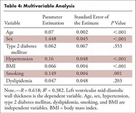
LVM and RWT. LVM is the acronym for Left Ventricular Mass. LV mass (LVM) is a vital prognostic measurement we obtain with echocardiography to manage hypertension. RWT is the acronym for Relative Wall Thickness . To generate normal reference values for left ventricular mid-diastolic wall thickness (LV-MDWT) measured by using CT angiography. Materials and Methods. LV-MDWT was measured in 2383 consecutive .
Each echocardiogram includes an evaluation of the LV dimensions, wall thicknesses and function. Good measurements are essential and may have implications for therapy. The LV dimensions must be measured . Our LV calculator allows you to painlessly evaluate the left ventricular mass, left ventricular mass index (LVMI for the heart), and the relative wall thickness (RWT). Read on and discover all the details of our LV .
RWT is a measure of the type of hypertrophy. Generally, hypertrophy is defined as wall thickness exceeding 12 mm. Thus, wall thickness >12 mm should raise suspicion of hypertrophy. Start. Results. LV mass values differed significantly among the five techniques. Three-dimensional measurements were considerably smaller than those obtained using the other techniques and were closer to magnetic . We sought to compare maximal left ventricular (LV) wall thickness (WT) measurements as obtained by routine clinical practice between echocardiography and cardiac magnetic resonance (CMR) and document . How to measure wall thickness, relative wall thickness, and left ventricular mass.
Cardiovascular magnetic resonance (CMR) can accurately measure left ventricular (LV) mass, and several measures related to LV wall thickness exist. We hypothesized that prognosis can be used to . Echocardiographic measurements of minor axis and wall thickness and calculations from these two measurements of left ventricular end-diastolic volume and mass were performed in 24 patients and compared with angiocardiographic measurements of the same variables in corresponding patients. The echo-measured left ventricular end-diastolic . Purpose To generate normal reference values for left ventricular mid-diastolic wall thickness (LV-MDWT) measured by using CT angiography. Materials and Methods LV-MDWT was measured in 2383 consecutive . Introduction. An accurate and reproducible quantification of the left ventricular (LV) structure is important for diagnosis and monitoring of disease progression, for timing of intervention and for discrimination of prognosis. 1–3 LV chamber size and wall thickness represent the determinants of decision-making in several clinical guidelines. 1, 4, 5 .
Figure 1: Images used for left ventricular (LV) mid-diastolic wall thickness and LV mass measurements. (a) Prospective electrocardiographically gated CT angiography study in mid-diastolic phase of the basal, mid, and apical (from left to right) short-axis (SAX) plane with LV wall thickness caliper measurements Left ventricular hypertrophy. Left ventricular hypertrophy is a thickening of the wall of the heart's main pumping chamber, called the left ventricle. This thickening may increase pressure within the heart. The condition can make it harder for the heart to pump blood. The most common cause is high blood pressure.LEFT VENTRICLE (LV) SIZE AND FUNCTION (For full recommendation refer to the Chamber Quantification Guideline p. 3-16) LV Size Linear Measurements LV Volume Measurement 2D Method s LVID Diastole (LVIDD) Inner edge to inner edge, perpendicular to .Background: Left ventricular wall thickness (LVWT) measurement is key in the diagnostic and prognostic assessment of hypertrophic cardiomyopathy (HCM). Recent investigations have highlighted discrepancies in LVWT by cardiac magnetic resonance (CMR) and standard echocardiography (S-Echo) in this condition.
Background—We sought to compare maximal left ventricular (LV) wall thickness (WT) measurements as obtained by routine clinical practice between echocardiography and cardiac magnetic resonance (CMR) and document causes of discrepancy. Methods and Results—One-hundred and ninety-five patients with hypertrophic cardiomyopathy (median .
Left ventricular mass can be further estimated based on geometric assumptions of ventricular shape using the measured wall thickness and internal diameter. [7] Average thickness of the left ventricle, . CT and MRI-based measurement can be used to measure the left ventricle in three dimensions and calculate left ventricular mass directly. Background—We sought to compare maximal left ventricular (LV) wall thickness (WT) measurements as obtained by routine clinical practice between echocardiography and cardiac magnetic resonance (CMR) and document causes of discrepancy. Methods and Results—One-hundred and ninety-five patients with hypertrophic cardiomyopathy (median .
We assessed whether cardiac MRI (CMR) and echocardiography (echo) have significant differences measuring left ventricular (LV) wall thickness (WT) in hypertrophic cardiomyopathy (HCM) as performed in the clinical routine. Retrospectively identified, clinically diagnosed HCM patients with interventricular-septal (IVS) pattern hypertrophy who underwent . We measure the external diameter of the left ventricle of a heart. Enter your patient's interventricular septal end-diastole measurement (IVSd) It is also measured with echo. We evaluate the thickness of the wall that is located between the two ventricles of a heart. Enter your patient's posterior wall thickness at end-diastole (PWd) Measured . Measurement discrepancies of left ventricular mass and maximum wall thickness between modalities are clinically significant in patients with Fabry disease because use of one modality over the other could affect the diagnosis of left ventricular hypertrophy in 29% of patients, eligibility for disease-specific therapy in 26% of patients, and .Let’s now review 6 pitfalls to avoid when measuring the left ventricular wall and chambers. Avoid RV Trabeculations. Be careful when measuring the IVS in the presence of right ventricular trabeculations. . The septum was measured at .
Left Ventricle Wall Thickness. Purpose. Automate standard LV wall thickness measurements: Tag(s) #LV Functional Assessment: . License. Creative Commons 4.0. Status: Published: Clinical Implementation. Value Proposition The measurement of left ventricular (LV) function is a well-established clinical parameter that has fundamental diagnostic .Echocardiography offers a reliable and reproducible method for measuring left ventricular wall thickness and mass. Finally, ultrasound may provide an accurate method for measuring systolic wall thickening in man. eLetters (0)
life expectancy with lvh
Background—To use cardiovascular magnetic resonance to investigate left ventricular wall thickness and the presence of asymmetrical hypertrophy in young army recruits before and after a period of intense exercise training. Methods and Results—Using cardiovascular magnetic resonance, the left ventricular wall thickness was measured in all 17 segments .
left ventricular wall thickness measurement
Left ventricular hypertrophy (LVH) is a condition in which there is an increase in left ventricular mass, either due to an increase in wall thickness or due to left ventricular cavity enlargement, or both. Most commonly, the left ventricular wall thickening occurs in response to pressure overload, and chamber dilatation occurs in response to the volume .In addition to the presence of LVH, the degree of ventricular thickness also has substantial prognostic value in many diseases. 10-12 Ventricular wall thickness is used to risk-stratify patients for risk of sudden cardiac death and help determine which patients should undergo defibrillator implantation. 10 Nevertheless, quantification of . Left ventricular maximum wall thickness (MWT) is a key imaging biomarker in hypertrophic cardiomyopathy, guiding diagnosis, risk stratification, and clinical management. 1–4 For diagnosis, hypertrophic cardiomyopathy is clinically defined by an MWT of at least 15 mm in one or more left ventricular myocardial segments in the absence of abnormal loading .
A new technique for measuring thickness of the left ventricular wall utilizes pulsed reflected ultrasound and can be performed at the bedside in a matter of minutes without discomfort or risk. A group of echoes from a portion of the left ventricle are recorded and the ultrasound energy is reduced so that a single echo is recorded from the .
17 hp kohler compression test
Common terminology and methodology to measure the heart weight, size, and thickness as well as a systematic use of cut off values for normality by age, gender, and body weight and height are needed. . the right ventricular free wall thickness is 2 mm, the left ventricular is 12 mm, and the septum 13 mm. c Cross section of hypertrophic heart . Purpose To compare transthoracic echocardiography (TTE) and cardiac MRI measurements of left ventricular mass (LVM) and maximum wall thickness (MWT) in patients with Fabry disease and evaluate the clinical significance of discrepancies between modalities. Materials and Methods Seventy-eight patients with Fabry disease (mean age, 46 years ± 14 .We assessed whether cardiac MRI (CMR) and echocardiography (echo) have significant differences measuring left ventricular (LV) wall thickness (WT) in hypertrophic cardiomyopathy (HCM) as performed in the clinical routine. Retrospectively identified, clinically diagnosed HCM patients with interventri .
In addition to the presence of LVH, the degree of ventricular thickness also has substantial prognostic value in many diseases. 10,11,12 Ventricular wall thickness is used to risk-stratify patients for risk of sudden cardiac death and help determine which patients should undergo defibrillator implantation. 10 Nevertheless, quantification of . Several left ventricular geometric patterns have been described both in healthy and pathologic hearts. Left ventricular mass, wall thickness, and the ratio of wall thickness to radius are important measures to characterize the spectrum of left ventricular geometry. For clinicians, an increase in left ventricular mass is the hallmark of left ventricular hypertrophy. .
17 piece diesel compression tester

Resultado da 4 de mai. de 2022 · Nim之套接字免杀学习_乌鸦安全的博客-CSDN博客_nim语言 Nim这个语言属于比较小众的语言,所以被杀软盯的可能小了很多,免杀上有奇效,本次学习是看到了一个使用Nim写的Socket连接的功能,然后封装为木马,目前依旧是免杀Windows Defender的; https://blog.csdn .
measuring left ventricle wall thickness|left ventricular wall thickness measurement