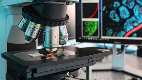
new laboratory technology used for analyzing the cell|advances in cell biology : exporters Fluorescence lifetime imaging microscopy, or FLIM, is a commonly used biomedical imaging technique that looks for the telltale light signatures of specific molecules. That information can illuminate what’s happening in and . As of version v2.0042, the seed used to determine the outcome of casting a spell is stored in your save file. More specifically, the result of casting any spell is based on your game seed (this is changed only on ascension) and your . Ver mais
{plog:ftitle_list}
web3 dias atrás · Previsão do tempo Iguaba Grande - RJ. 15:00 Terça. Períodos nublados 29° Sensação de 32° Este 22 - 39 km/h. Baixa pressão deixa a Região Sul sob alerta de .
new technology in imaging
iphone x vs note 8 drop test
microscopic cell biology research
The new approach makes use of green or red fluorescent molecules that flicker on and off at different rates. By imaging a cell over several seconds, minutes, or hours, and then extracting each of the fluorescent . Fluorescence lifetime imaging microscopy, or FLIM, is a commonly used biomedical imaging technique that looks for the telltale light signatures of specific molecules. That information can illuminate what’s happening in and .Enhance crop management and optimize yields with NEW AGE Laboratories' plant sap analysis services. Gain real-time insights into nutrient availability and plant health to make informed adjustments to amendments and additives. Our .
As in all experimental sciences, research in cell biology depends on the laboratory methods that can be used to study cell structure and function. Many important advances in understanding cells have directly followed the development of new methods that have opened novel avenues of investigation. An appreciation of the experimental tools available to the cell biologist is thus . Now, researchers at The Jackson Laboratory (JAX) have combined advanced imaging techniques with a new computational method to probe how immune cells interact with each other in never-before-seen .The Laboratory for Cell Analysis (LCA) was established in 1983 with the following responsibilities: Provide flow and image cytometers and the technical support needed to make use of these instruments. Educate UCSF students, staff, and faculty in cytometry technology and applications. Develop new cytometric methods and new cytometric applications.

As the new year begins, clinical laboratory managers must proactively anticipate the emerging trends that will redefine the healthcare landscape in 2024. One of the key focal points revolves around integrating emerging technologies (ETs) into laboratory medicine, a trajectory set to profoundly transform diagnostics and prevention.Stem cell-based therapies are defined as any treatment for a disease or a medical condition that fundamentally involves the use of any type of viable human stem cells including embryonic stem cells (ESCs), iPSCs and adult stem cells for autologous and allogeneic therapies . Stem cells offer the perfect solution when there is a need for tissue .
iphone x vs s8 plus drop test
Digital transformation with new laboratory technology helps reduce errors, speed up lab functions and optimize data handling. . Cell counting, for example, can be a rigorous procedure in cell culture labs . Traditionally, scientists have calculated cell concentrations and carried out viability assays manually with a grided hemocytometer . There are various laboratory technologies used to analyze different types of cells. There are new laboratory technologies,which make scientists understand cells and their unique attributes better. New cell technologies enable scientists uncover more information about how cells function and how they interact with each other. This paper will . In recent years, stem cell therapy has become a very promising and advanced scientific research topic. The development of treatment methods has evoked great expectations. This paper is a review focused on the discovery of different stem cells and the potential therapies based on these cells. The genesis of stem cells is followed by laboratory steps of controlled . 1 INTRODUCTION. Flow cytometry is a powerful technology used to analyze and measure the physical, chemical, and/or gene expression characteristics of cells or particles. 1 This technique allows clinical laboratories and researchers to measure numerous properties of individual cells in a rapid, multiplexed, and quantitative manner. Given the high flexibility of .

blood analysis, laboratory examination of a sample of blood used to obtain information about its physical and chemical properties. Blood analysis is commonly carried out on a sample of blood drawn from the vein of the arm, the finger, or the earlobe; in some cases, the blood cells of the bone marrow may also be examined. Hundreds of hematological tests and . With the rapid development of high-throughput technology 6, several new technologies are widely used in proteomics and metabolomics in recent years.Regardless the specific technique, these global . Figure 1: View of the low angle light scatter x axial light loss histogram. Different particles that cause cellular interference in the WBC counting have a distinct signature pattern in this new histogram. 1a: nRBCs typically plot in the center of the histogram and their population is highlighted in red. 1b: Giant platelets form a vertically stretched green population on the left .
Test and analyze body fluids, such as blood, urine, and tissue samples; Operate laboratory equipment, such as microscopes and automated cell counters; Use automated equipment that analyzes multiple samples at the same time; Record data from medical tests and enter results into a patient’s medical record
New automation and digital tools could sense and respond to changing growth conditions, delivering cells to patients faster and at lower cost than ever before.
Clinical laboratories are healthcare facilities providing a wide range of laboratory procedures that aid clinicians in diagnosing, treating, and managing patients.[1] These laboratories are manned by scientists trained to perform and analyze tests on samples of biological specimens collected from patients.
Automation and how it can benefit the lab The traditional use of automation in the laboratory was mainly for pharmaceutical assays. It helped with drug synthesis on a large scale and high-throughput analysis of .
Genetic testing is the laboratory analysis of human chromosomes, DNA and RNA to detect genetic material and/or identify genetic changes. . The ongoing development of new gene sequencing technology and the declining cost of sequencing has led to the development of tests that can look for genetic disorders beyond a single gene. The following . Bond et al. provide a technologically focused review on common super-resolution light microscopy approaches as well as recent, promising technical developments in this field. The review particularly highlights emerging advanced imaging approaches that hold promise for visualizing the molecular architecture of cells with high spatiotemporal resolution. Single-cell transcriptomics was also used in kidney organoids to assess cell maturation and fate trajectories, to determine the effects of morphogens, to improve protocols, and to analyze disease phenotypes. Comparative analysis of human kidney organoids revealed remarkable consistency with podocytes in the fetal human kidney (Tran et al., 2019). Some of the methods laboratory professionals use to analyze samples have been in use for over a century. One such staple, the Gram stain, was introduced in 1882. It uses two different dyes and .
Founded at the Massachusetts Institute of Technology in 1899, MIT Technology Review is a world-renowned, independent media company whose insight, analysis, reviews, interviews and live events .
Bloodstain pattern analysis (BPA) software is an emerging technology in forensic science that seeks to reconstruct crime scenes by analyzing blood and bloodstains. This computer-based software can estimate the Area(s) of Origin (AO) of a bloodletting event, providing crucial insights into the sequence of events at a crime scene. Cryo-EM is just one of many imaging tools driving rapid advances in cell biology and related fields. “New techniques have come on board over the last ten years that allow us to see things inside .Hematology analyzers are used widely in patient and research settings to count and characterize blood cells for disease detection and monitoring. Basic analyzers return a complete blood count (CBC) with a three-part differential white blood cell (WBC) count. Sophisticated analyzers measure cell morphology and can detect small cell populations to diagnose rare blood .
Such technology can, in theory, incorporate all the key steps of molecular detection: cell lysis and extraction, nucleic acid purification, amplification and detection of reaction products. Such miniaturisation may also allow multiplexing to enable high-throughput testing within a single portable device [ 118 ].Conduct molecular analysis of a single cell type, including RNA expression and epigenetic analysis; Genetically modify and expand a particular cell type of interest for disease modelling or cell therapy research applications (e.g. T cell therapy research) Directly use purified cells for adoptive cell transfer experiments in various animal models

Resultado da Hours. See a problem? Let us know. Get directions, maps, and traffic for Ashfield-Colborne-Wawanosh. Check flight prices and hotel availability for your visit.
new laboratory technology used for analyzing the cell|advances in cell biology