tear duct test dog|lacrimal duct in dogs : sourcing In calves, multiple openings of the nasolacrimal duct may empty tears onto the lower eyelid and medial canthus, causing chronic dermatitis. Treatment in dogs, foals, and young camelids consists of surgically opening the blocked orifice .
Descubra o universo de jogos no LEOBET, a plataforma de cassino online definitiva, oferecendo uma vasta gama de jogos de apostas. Com uma operação licenciada pela .
{plog:ftitle_list}
webPreencher login com seu CPF e a senha são os ultimos 5 dígitos do CPF, clique em entrar. Acesse www.azza.net.br e clique em "Minha Conta" O QUE É A CENTRAL DO .


In some cases, the obstruction is related to the shape and size of the dog's head and muzzle. Obstruction may also be caused by a hereditary defect in the formation of the nasolacrimal duct. This defect results in the lack of an opening where the nasolacrimal duct meets the conjunctiva (pink tissue surrounding . See moreThe signs of nasolacrimal duct obstruction are largely cosmetic in nature. Most affected dogs have excessive watering of the eyes or reddish-colored tear staining of . See more
lacrimal duct obstruction dogs
Nasolacrimal duct obstruction is often diagnosed using a dye known as fluorescein. This dye fluoresces (glows) under a black light, allowing your veterinarian to . See moreIn cases of nasolacrimal duct obstruction caused by inflammation, anti-inflammatory medications and antibiotics may alleviate the obstruction. Use all . See moreWithout treatment, nasolacrimal duct obstruction will cause continued issues with tear staining. Untreated dogs also have an increased likelihood of skin . See moreIn calves, multiple openings of the nasolacrimal duct may empty tears onto the lower eyelid and medial canthus, causing chronic dermatitis. Treatment in dogs, foals, and young camelids consists of surgically opening the blocked orifice .

Nasolacrimal ducts allow tears to drain from each eye into the nose. Disorders of these structures can lead to either eyes that water excessively or dry eyes. They may be congenital (present at birth) or caused by infection, foreign objects in .
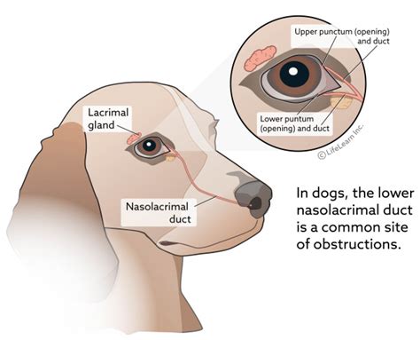
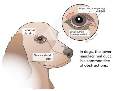
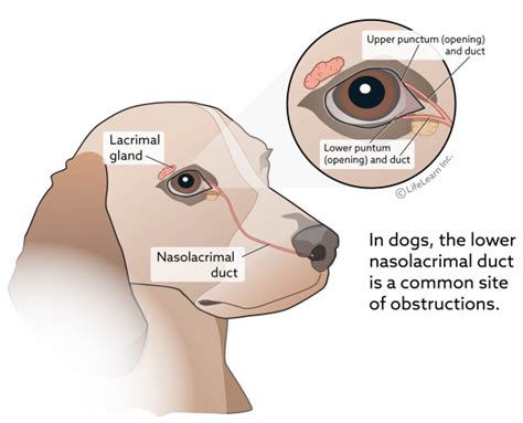
Note the relationship of the eyelids, puncta, canaliculi, lacrimal sac, nasolacrimal duct, and nasal puncta. Schirmer Tear Test. The STT should be the first diagnostic test completed during examination of a dog with epiphora.At a minimum, a Schirmer tear test (STT), corneal fluorescein staining (dying the cornea so your veterinarian can see any defects), and tonometry (checking eye pressure) is recommended. If .There are several treatments that can be used in dogs to unblock a tear duct and restore functioning. The most commonly used is to flush the tear duct to remove blockage, clear .The nasolacrimal duct terminates at the nasal punctum located on the ventrolateral floor of the nasal vestibule, approximately 1cm caudal to the external nares. Approximately 50 per cent of .
During the examination of your pet, we will test the patency of the eyelid punctae and nasolacrimal duct using a fluorescein green dye. If the dye fails to passively flow out the .One of the simplest tests to assess tear drainage is to place a drop of fluorescein stain in the eye, hold the dog’s head slightly downward, and watch for drainage into the nose. If the drainage system is functioning normally, the .
Your dog’s tear duct drains tears produced to lubricate the surface of the eye to the nose. It is referred to as the naso-lacrimal duct and carries tears through the bones into the nasal passage. Tears are required to continuously flush the eye surface of bacteria and debris. If the naso-lacrimal duct becomes blocked, does not open properly . Dogs — Tear duct malformations and excess tear spillover are common in dogs, but dacryocystitis occurs less frequently. Tear staining is most often observed in brachycephalic (i.e., flat-faced) breeds with large eyes and . See your doctor if your symptoms worsen. Physical examination by a medical professional is required in order to diagnose a blocked tear duct. While simple inflammation might be causing the blockage, it could also be a tumor or .
A swollen tear duct can be caused by a blockage or infection. It can be treated with self-care, medication, or surgery. . Tear drainage test: This test measures how quickly your tears are draining. One drop of a special dye is placed on the surface of each eye. If the drop is still on the surface of the eye after five minutes, this could . For most dogs, epiphora and subsequent tear staining is caused by variations in anatomy that cause tears to drain onto the face, rather than into the nose via the nasolacrimal duct. In some dogs . The nasolacrimal duct system of the dog is similar to that of most domestic mammals. It is a walled conduit that drains the tear film from the eye into the nasal passages. . Note the relationship of the eyelids, puncta, canaliculi, lacrimal sac, nasolacrimal duct, and nasal puncta. Schirmer Tear Test. The STT should be the first diagnostic .Fluorescein Dye Test helps in determining the extent of dryness and assess the impact on the cornea. Nasolacrimal Duct Blockage (NLD Blockage)- Jones Test: Pets suffering from epiphora or tear streak problem may be having the blockage of NLD and FDT is the simplest test to perform which can help in ruling out it. In case of a patent NLD, dye is .
The tear ducts drain tears into the back of the sinuses and down the throat. Epiphora can be caused by either insufficient drainage of tears through the tear ducts, or by an excessive production of tears. . Some of the causes of increased tear production in dogs include conjunctivitis (viral or bacterial), allergies, eye injuries, abnormal . A blocked tear duct means tear fluid can’t flow out of your eye properly. It’s common in babies but can happen in adults. It’s usually very treatable. . One simple test they can do is called the “dye disappearance test.” To do it, a provider adds a drop of a special dye called fluorescein to your eye. Fluorescein glows under a blue . Tests used to diagnose a blocked tear duct include: Tear drainage test. This test measures how quickly your tears are draining. One drop of a special dye is placed on the surface of each eye. You may have a blocked tear duct if after five minutes most of the dye is still on the surface of your eye. Irrigation and probing.All owners need to know about dry eye in dogs - including symptoms, treatment and breeds prone to dry eye. Donate Menu. Pet help & advice; . Your vet can diagnose dry eye based on your dog’s symptoms and by performing a ‘Schirmer Tear Test’ (STT). . you will need to consider options such as a parotid duct transposition or even .
This simple test uses a special wicking paper to measure the amount of tear film produced in one minute. Additional diagnostic tests may include corneal staining to check for corneal ulcers, intraocular pressure (IOP) to determine if glaucoma is present, and tear duct examination or flushing to ensure normal tear drainage. How is dry eye treated?
This new route bypasses the duct that empties into your nose (nasolacrimal duct), which is typically the blockage site. Stents or intubation typically are placed in the new route while it heals, and then removed three or four months after surgery. The steps in this procedure will vary depending on your particular tear duct blockage. For a few hours after tear duct probing, some children have blood-colored fluid drain from the eye. Using antibiotic eye drops or ointment a few times a day for about a week can help prevent an .
The quantity of tears is measured using a Schirmer Tear Test strip. These are placed on the eye. Normal tear production in the dog is 15-25mm/min. Dry eye is usually treated with a combination of topical lubricants and tear stimulants .Objective: To evaluate fluorescein nasolacrimal transit (NLT) times in ophthalmically normal dogs and nonbrachycephalic cats by use of 2 methods of the Jones test. Animals: 73 dogs and 36 cats. Procedures: Fluorescein dye was applied to the ocular surface of both eyes by means of a wetted fluorescein strip and, in a subsequent test, by administration of a drop of 0.2% .
lacrimal duct in dogs treatment
This veterinarian-reviewed article explains that blocked tear ducts in dogs have a variety of causes, including eye infection, scar tissue, swelling, or a lack of tear duct development. Excess tears, staining around the eyes, and .In dogs the disorder is believed to be caused by an autoimmune disease in which the dog’s own immune system attacks and destroys the tear producing glands around the eye. The dog’s body “reads” the production of tears from the tear ducts as invading enemy bacteria and attacks the area, as it would with any other infection. The most important thing is to make sure that your dog is healthy and his eyes don’t tear up more than necessary. If his eyes water more than normal, it could be a sign that he has an allergy, that his tear ducts are blocked, or that he has an eye infection.. Normally this comes along with red eyes, but even without the redness, tearing up too much could be a sign .
The precorneal tear film is a substantial structure both in its size and functional importance. Yet it's difficult to directly evaluate. Various studies have come up with imaging techniques to measure the thickness of the tear film and although one study measured it at a whopping 40um, essentially equaling the size of the epithelium, most still feel it sits .Objective: The present study aimed to determine the effects of age, sex, reproductive status, skull type, and nasolacrimal duct (NLD) patency on tear production and tear film breakup time (TBUT) in normal dogs. Animals studied: The ophthalmic data of 82 healthy adult dogs were evaluated in this study. Procedures: Age, sex, breed, and reproductive status were recorded.

For dog owners of long-haired or wrinkly breeds such as English Bulldogs, Frenchies, and Pugs, blocked tear ducts can be a troublesome issue. Blocked tear ducts in dogs are common among these breeds and can cause excessive tearing, irritation, and infections if left untreated. In this blog post, we'll discuss the causes and symptoms of blocked tear . Obstruction of the Tear Duct. In healthy dogs, the normal tear secretion is drained through the tear ducts that are at the inner corner of the eye. When the tear ducts are partially or obstructed, epiphora will occur, and the tears will run down your dog’s face, staining the area around the eyes because they have nowhere else to drain. The tear stains can become worse when pup is teething and sometimes clears up afterwards. My son's dog is totally clear of tear stains now at 4 years old. My dog still has them but not as much as she used to but I do wipe around the under eye area every day with Optrex Multi Action Eye Wash which helps keep them down. When she was spayed at 10 . What causes a dog’s tear duct to swell? “Cherry eye,” as it is commonly referred to, is a prolapsed gland of the nictitans. . Occasionally, a modified fluorescein dye disappearance test may be done. See also Are dogs allowed at El Dorado State Park? Do tear ducts dry up? Dry eye can be temporary or chronic. It occurs when your tear .
A blockage in your tear duct causes dacryocystitis. These blockages disrupt the flow of tears from your eyes into your nasal cavity. A membrane that blocks the duct causes the blockage in newborns. In children and older people, blockages can be caused by many things. Lacrimal and Third Eyelid Glands. The gland of the third eyelid, which lies within the stroma of the third eyelid, is partially visible on the inner surface of the third eyelid (see Chapter 8 and Figure 9-2).The tubuloalveolar lacrimal gland is flattened and lies over the superior-temporal part of the globe. In the dog it lies beneath the orbital ligament and supraorbital .
top en 960 hail impact tester
lacrimal duct in dogs
Streaming filmów online za darmo zrewolucjonizował sposób, w jaki oglądamy filmy. Dzięki wygodzie platform do przesyłania strumieniowego online możesz uzyskać dostęp do ogromnej https://movieflix.pl/ biblioteki filmów w zaciszu własnego domu. Chociaż istnieją korzyści, takie jak opłacalność i różnorodność treści, istnieją .
tear duct test dog|lacrimal duct in dogs