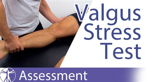
test for medial collateral ligament tear|medial collateral ligament test knee : solution A medial collateral ligament (MCL) injury is a stretch, partial tear, or complete tear of the ligament on the inside of the knee. A valgus trauma or external tibia rotation are the causes of this injury.
WEBPlay the Fire Dozen demo by CT Gaming play for Free Slot Review ️ Check the complete list of casinos where you can play for real January 2023 ️
{plog:ftitle_list}
WEBLas Vegas.C$285 par passager.Départ le lun. 25 mars, retour le mer. 1 mai.Vol aller-retour avec Lynx Air.Vol aller direct avec Lynx Air, aéroport de départ Montréal Pierre Elliott Trudeau, partant le lun. 25 mars, arrivant à Harry Reid International de Las Vegas.Vol retour direct avec Lynx Air, au départ de Harry Reid International de Las .
windows 10 memory hard drive test
valgus stress test positive result
The valgus stress test, also known as the medial stress test, is used to assess the integrity of the medial collateral ligament (MCL) of the knee. MCL injuries are common in the athletic population and can occur as either isolated injuries, or combined with other structural injuries . See moreOverall, the MCL plays a crucial role in the stability of the knee and acts as the primary valgus restraint in a flexed knee. If knee hypermobility is present due to a sprained MCL, the ACL is placed under higher stress loads with valgus forces (particularly at 45 . See more An MCL tear is damage to the medial collateral ligament, which is a major ligament that’s located on the inner side of your knee. The tear can be partial (some fibers in the .
special tests for mcl tear
A medial knee ligament sprains (MCL) is a tear of the ligament on the inside of the knee. In order to diagnose an MCL sprain your physio or doctor performs a number of tests including the valgus stress test. Knee ligament .
Damage to your medial collateral ligament (MCL) is called an MCL tear. A tear can be either partial or complete. When some fibers in the ligament are torn, it is a partial tear.A medial collateral ligament (MCL) injury is a stretch, partial tear, or complete tear of the ligament on the inside of the knee. A valgus trauma or external tibia rotation are the causes of this injury.
A medial collateral ligament (MCL) knee injury is a traumatic knee injury that typically occurs as a result of a sudden valgus force to the lateral aspect of the knee. Diagnosis can be suspected with increased valgus laxity . The medial collateral ligament, or MCL, of the knee can tear due to injury and cause pain. Treatment depends on the severity of the injury. Learn more about MCL tears here.
The medial collateral ligament (MCL) is a flat band of connective tissue that runs from the medial epicondyle of the femur to the medial condyle of the tibia. Its role is to provide . Medial collateral ligament injury occurs when excessive valgus stresses or external rotation forces are placed on the knee joint. The most common symptom is medial . Collateral Ligaments. Valgus stress test for Medial Collateral Ligament. It is performed with the patient supine and the knee in 20° of flexion. With one hand on the lateral aspect of the knee and the other on the foot, the . Medial collateral ligament injuries of the knee: current treatment concepts. Curr Rev Musculoskelet Med. 2008 Jun;1(2):108-13. doi: 10.1007/s12178-007-9016-x. PMID: 19468882; PMCID: PMC2684213. .
The medial collateral ligament's main function is to prevent the leg from extending too far inward, but it also helps keep the knee stable and allows it to rotate. Injuries to the medial collateral ligament most often happen when the knee is hit directly on its outer side. The medial collateral ligament usually responds well to nonsurgical treatment.Valgus Stress Test: Medial collateral ligament injury (Joint line tenderness may also indicate meniscal tear) The patient is supine and the knee held by the examiner at 30° of flexion (or the patient sitting with the thigh on the couch and leg hanging over the edge at a 30° angle) with the examiner's hand holding the ankle or foot, and . The medial collateral ligament (MCL) is one of four major ligaments that are critical to the stability of the knee joint. A ligament is made of tough fibrous material and functions to control excessive motion by limiting joint mobility. Also has a deep attachment to the medial meniscus. Lateral collateral ligament (LCL) - prevents medial movement of the tibia on the femur when varus (towards the midline) stress is placed on the knee. Runs between the lateral epicondyle of the femur and the head of the fibula. Also known as the fibular collateral ligament (FCL).
The medial collateral ligament (MCL) is located on the inner aspect, or part, of your knee, outside the joint. Injury to the MCL is often called an MCL sprain or tear. MCL injuries are common in .There is often a positive valgus stress test. (Valgus pressure is applied to the knee in full extension and at 30° flexion - there is pain, laxity or medial joint gapping/opening in a positive test.) Lateral collateral ligament injury. A lateral collateral ligament injury is less common than a medial collateral ligament injury. A medial knee ligament sprains (MCL) is a tear of the ligament on the inside of the knee. In order to diagnose an MCL sprain your physio or doctor performs a number of tests including the valgus stress test. Knee ligament sprains are graded 1, 2, or 3 depending on the severity of the injury. Thyroid Test Analyzer; Doctor Discussion Guides; Hemoglobin A1c Test Analyzer; . The medial collateral ligament (MCL) on the inner side of the knee is most often torn when there is a force that strikes the outside of the knee. . Miyamoto RG, et al. Treatment of Medial Collateral Ligament Injuries. J. Am. Acad. Ortho. Surg., March 2009; 17: .
A block to the outside part of the knee during football is a common way for this ligament to be injured. An MCL injury can be a stretch, partial tear, or complete tear of the ligament. MCL injuries also often occur at the same time as an anterior cruciate ligament (ACL) injury. Common symptoms of an injury to the medial collateral ligament are:
The medial collateral ligament (MCL) is a flat band of connective tissue that runs from the medial epicondyle of the femur to the medial condyle of the tibia and is one of four major ligaments that supports the knee. MCL injuries often occur in sports, being the most common ligamentous injury of the knee, and 60% of skiing knee injuries involve the MCL).Surgery for medial collateral ligament injury. Most people recover from an MCL injury without needing to have surgery. But sometimes, surgery is the best option to repair an injury to the medial collateral ligament. This is most likely if: more than one ligament or tissue in your knee is damaged; your knee remains unstable after physiotherapy
Medial collateral ligament tears often occur as a result of a direct blow to the outside of the knee. This pushes the knee inward (toward the other knee). . Other tests that may help your doctor confirm your diagnosis include: X-rays. Although they will not show any injury to your collateral ligaments, X-rays can show whether the ligament .The medial collateral ligament, or MCL, is a broad, thick band that runs down the inner part of the knee, from the femur (thighbone) to a point 1.5 to 2 inches from the top of the tibia (shinbone). . In addition, your doctor may order the following tests: X-ray. This can show other damage and bone injury. Magnetic resonance imaging (MRI .The medial collateral ligament (MCL) is located on the inner side of your knee and connects the thigh bone (femur) and the shinbone (tibia). . but your doctor may also complete the following tests to see how bad the sprain or tear is: X-rays, which can help rule out further damage such as a broken or fractured bone. Magnetic resonance imaging .Purpose: The Valgus Stress Test is used to assess the integrity of the MCL or medial collateral ligament of the knee.. How to Perform Valgus Stress Test. Position of Patient: The patient should be relaxed in the supine position. .
ONLINE COURSES: https://study.physiotutors.comGET OUR ASSESSMENT BOOK ︎ ︎ http://bit.ly/GETPT ︎ ︎OUR APPS: 📱 iPhone/iPad: https://apple.co/35vt8Vx🤖 Andro. - Pivot shift test A; Pictures - Medial knee structures to palpate - Posterior intra-articular view of knee joint . Injuries of the medial collateral ligament (MCL), also referred to as the tibial collateral ligament, occur frequently in athletes, particularly those involved in sports that require sudden changes in direction and speed, and in .
positive valgus stress test knee
A medial collateral ligament tear will typically occurs after trauma to the lateral aspect of the knee. . The main clinical finding on examination will be increased laxity when testing the MCL*, via the valgus stress test. The patient will be extremely tender along the medial joint line, but may be able to weight bear. .
medial collateral ligament test knee
Medial or lateral knee pain with corresponding joint-line tenderness can result from acute injury or chronic overuse and may indicate meniscal derangement or a sprain or rupture of a collateral . Tears of the medial collateral ligament (MCL) are the most common knee ligament injury. Incomplete tears (grade I, II) and isolated tears (grade III) of the MCL without valgus instability can be treated without surgery, with early functional rehabilitation. . This test determines whether the injury primarily affects the sMCL or the POL and dMCL.Of the collateral ligament injuries, MCL injuries are more commonly . Tenderness and pain at the lateral side of the knee and at the injury; The varus test shows a significant joint laxity (>10mm laxity) . in a hinged-brace for the following 3 to 6 weeks while performing functional rehabilitation in order to maintain medial and lateral .
The lateral collateral ligament (LCL), also known as the fibular ligament, is one of the knee joint's key stabilizers (see Image. Left Knee Ligaments). This fibrous structure originates from the lateral femoral epicondyle and inserts on the fibular head. The LCL is part of the knee's "posterolateral corner" (PLC) along with the biceps femoris tendon and fibular .
windows 10 please run hard disk test
http://www.johngibbonsbodymaster.co.uk/courses/John Gibbons is a sports Osteopath and a lecturer for the 'Bodymaster Method ®' and in this video he is demons.
windows 10 run hard drive test

web3 de nov. de 2019 · Concordo com o Bruno! Contas universitárias se distinguem das outras pela gratuidade ou pelas taxas menores (limites menores também). O Nubank não .
test for medial collateral ligament tear|medial collateral ligament test knee