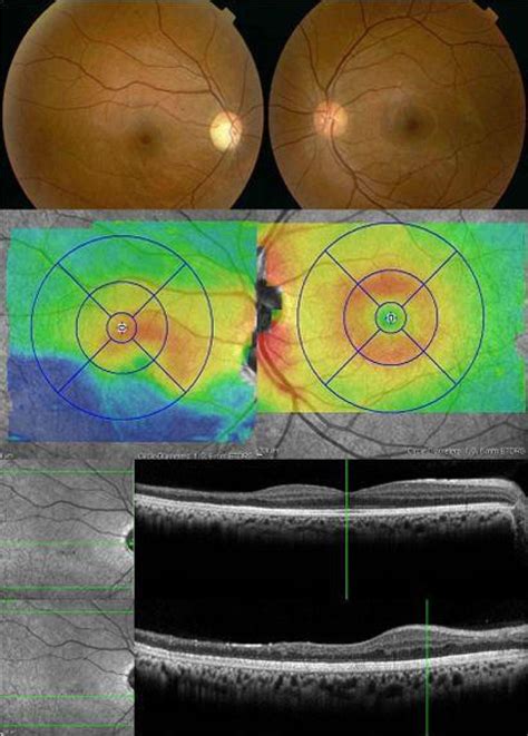
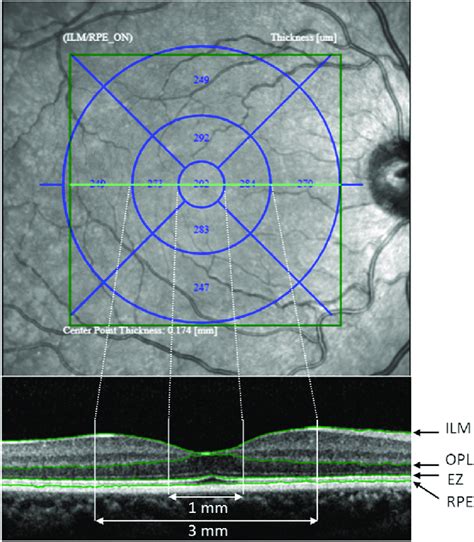
thickness measurement of retinal layers|full thickness retinal hole : trade The thickness values of each retinal layer in a large white population are provided. The thickness of retinal layers is influenced by gender, sex, and AXL, with a variable extent depending on the analyzed ETDRS map ring. Publisher: monotool. Release date: 10 Nov, 2023. Version: Final. Language: English (MTL) Censored: Yes (Mosaics) Store: DLsite. OtomiCloud – MegaUp – WorkUpload – .
{plog:ftitle_list}
Marcahuasi is a plateau in the Andes Mountains, located 60 km east of Lima, on the mountain range that rises to the right bank of the Rímac River. The site is located at 4,000 metres (13,000 ft) above sea level and is known for its unusual geological formations; curious shapes of human faces and animals visible in granite rock.
We first calculated the mean and standard deviation of the main outcome parameters. i.e. the thickness of the retinal layers in the fovea, parafovea and perifovea areas. Secondly, we compared the thickness of the . We developed the ThicknessTool (TT), an automated thickness measurement plugin for the ImageJ platform. To calibrate TT, we created a calibration dataset of mock .The thickness values of each retinal layer in a large white population are provided. The thickness of retinal layers is influenced by gender, sex, and AXL, with a variable extent depending on the analyzed ETDRS map ring.
Thickness measurement of retinal layers is important as it provides useful information for detecting pathological changes and diagnosing retinal diseases. Recently various approaches .
what is normal retinal thickness
Regional differences in the thickness of retinal layers. A, Papillomacular bundle, which has the thickest ganglion cell layer. B, Macula with 2-cell-thick ganglion cell layer. C, . Individual retinal layer thicknesses of five subfields in the macula were measured using automated retinal segmentation software packaged with the spectral-domain optical coherence tomography and. We evaluated the intra-retinal layer thickness alterations in 71 DM eyes with no diabetic retinopathy (DR), 90 with mild DR, and 63 with moderate DR without macular .The algorithm identified seven boundaries and measured thickness of six retinal layers: nerve fiber layer, inner plexiform layer and ganglion cell layer, inner nuclear layer, outer plexiform layer, outer nuclear layer and photoreceptor .
The mean thickness of the whole retina, outer plexiform layer, outer nuclear layer,retinal pigment epithelium, inner retinal layer and photoreceptor layer was 259.8 ± 18.9 . Thickness measurements of retinal layers derived from OCT images have potential value for objectively documenting disease-related retinal thickness abnormalities and .
Retinal thickness assessment has been one of the main applications of OCT since this technology was introduced into clinical practice. All commercially available posterior segment OCTs can provide retinal thickness maps based .If accurate measurements of retinal layers or their thickness proportions are of interest, the automatically calculated thickness values can either be corrected by manually adjusting the delineation for the retinal pigment epithelium or by subtracting a correction factor for total retinal thickness measurements, as described in Table 3. These .
Four SD-OCT measurements of outer retinal layer thickness were selected for our analyses of outer-retinal layer related boundaries as represented in Fig. 1: inner nuclear layer -retinal pigment .Purpose: To report an image segmentation algorithm that was developed to provide quantitative thickness measurement of six retinal layers in optical coherence tomography (OCT) images. Design: Prospective cross-sectional study. Methods: Imaging was performed with time- and spectral-domain OCT instruments in 15 and 10 normal healthy subjects, respectively.
This study aimed to develop a non-invasive diagnostic test for CI based upon retinal thickness measurements explored in a mouse model. Discrimination indices and retinal layer thickness of healthy . Accurate assessment of the retinal nerve fibre layer (RNFL) is central to the diagnosis and follow-up of glaucoma. The in vivo measurement of RNFL thickness by a variety of digital imaging . The exclusion criteria were as follows: BCVA <20 / 25; IOP ≥21 mmHg; a medical history of any systemic disease such as diabetes or hypertension; any ophthalmic disease that could affect the retinal layer thickness such as glaucoma and retinal and neuro-ophthalmic diseases; structural change caused by high myopia, including staphyloma, lacquer .
Optical coherence tomography (OCT) has been applied to measure peripapillary retinal nerve fiber layer (RNFL) thickness at a micrometer scale in several optic neuropathies such as non-arteritic .
The ability to segment retinal layers allows for thickness measurement, which improves glaucoma diagnosis, because thinning of the nerve fiber layer marks the onset and progression of the disease. . The precision of the placement of the follow up scans has been evaluated by means of retinal thickness measurements on FUP examinations. A .Measurement of retinal nerve fibre layer (RNFL) thickness in ODE patients may help in monitoring the progress of the disease and treatment response. Objective: To assess the clinical characteristics, aetiology and retinal nerve fibre layer (RNFL) imaging features of optic disc oedema (ODE) patients. Design: A retrospective observational study .This program was used to profile and measure the thickness of individual retinal layers, and layer combinations . 5-7 The segmentation approach is comparable to the software developed by Hood et al (MATLAB based; MathWorks; Natick, MA). 8 The following layer and layer combination thicknesses were measured: total retinal thickness (TR), retinal .
Purpose To clarify the abilities of circumpapillary retinal nerve fiber layer thickness (cpRNFLT) obtained by optical coherence tomography (OCT) and circumpapillary vessel density (cpVD) measured by OCT-angiography to distinguish different stages in primary open-angle glaucoma determined by 24–2 or 30–2 static visual field (VF) testing. Methods This . Current descriptions of retinal thickness across normal age cohorts are mostly limited to global analyses, thus overlooking spatial variation across the retina and limiting spatial analyses of . Optical coherence tomography (OCT) is used worldwide by clinicians to evaluate macular and retinal nerve fiber layer (RNFL) characteristics. It is frequently utilized to assess disease severity, progression and efficacy of treatment, and therefore must be reliable and reproducible. To examine the influence of signal strength on macular thickness parameters, .Objective: To measure the retinal nerve fiber layer (RNFL) thickness using optical coherence tomography (OCT) in optic atrophy eyes of patients with optic neuritis and investigate the correlation between the RNFL thickness and the visual function. Methods: To compare the RNFL thickness using StratusOCT, three groups of the subjects were enrolled, including 72 patients .
Similar misalignment with wrong measurements can happen while measuring central corneal thickness and peripapillary retinal nerve fiber layer thickness. In such cases, the image should be recaptured with proper positioning of the .
Vessel density may be more sensitive than retinal layer thickness measurement in the detection of inner retinal change in eyes with GA. View. Show abstract.

morphology of the retina at the level of individual retinal layers.5 As a result, alterations in single retinal layers occurring in specific diseases have been studied, often proving to be more reliable information than full retinal thickness measurements in predicting clinical outcomes and in understanding the pathophysiology of retinal . Various OCT devices have shown high reproducibility in distinguishing each layer of the retina in detail and accurately presenting the thickness of the entire retina, including the thickness of each layer 3, 4. Retinal thickness is also useful for evaluating the course of disease in conditions that cause macular edema, such as diabetic macular . Imaging and quantitative assessment of the retinal tissue is of value for the diagnosis of many retinal diseases. Optical coherence tomography (OCT) is an imaging technique that generates high-resolution cross-sectional images of the retina and provides quantitative measurement of the total retinal thickness. 1, 2 OCT has been extensively .
how hard is the ksnsas concealed carry test
how hard is the ky permit test
Spectral-domain OCT (SD-OCT) provides high resolution images enabling identification of individual retinal layers. We included 32,923 participants aged 40–69 years old from UK Biobank.DOI: 10.1016/J.AJO.2005.01.012 Corpus ID: 2962706; Quantitative thickness measurement of retinal layers imaged by optical coherence tomography. @article{Shahidi2005QuantitativeTM, title={Quantitative thickness measurement of retinal layers imaged by optical coherence tomography.}, author={Mahnaz Shahidi and Zhangwei Wang and Ruth Zelkha}, .
Thickness measurements of retinal layers derived from OCT images have potential value for objectively documenting disease-related retinal thickness abnormalities and monitoring progressive changes .
OCT images that observe the structure of the retina may di˜er in their retinal thickness measurements due to variations in the wavelengths used by the devices and image processing algorithms 5–8 . Hence, this study was conducted to measure the macular thickness, retinal nerve fiber layer thickness, and ganglion cell complex thickness using optical coherence tomography and to compare the macular thickness, retinal nerve fiber layer thickness, and ganglion cell complex thickness with that of normal controls among type 2 diabetics (with and .
To assess retinal nerve fiber layer (RNFL) thickness measurements of normal Northern Nigerian adults using optical coherence tomography (OCT).The OCT procedure was carried out with the Carl Zeiss Stratus OCT Model 3000 software version 4.0 (Carl Zeiss . Evaluation of Retinal Layer Thickness Parameters as Biomarkers in a Real-World Multiple Sclerosis Cohort. . Schmidt-Erfurth U, Reitner A. Effectiveness of averaging strategies to reduce variance in retinal nerve fibre layer thickness measurements using spectral-domain optical coherence tomography. Graefes Arch Clin Exp Ophthalmol. 2013; 251 .
retinal thickness treatment
retinal thickness problems

16 de nov. de 2018 · Download Simple LRC Format Lyrics which is the Music Subtitles of : AUD 20181116 WA0000; Length: 03:24.41 ; You can Download LRC, Copy and Edit the .
thickness measurement of retinal layers|full thickness retinal hole