confocal microscopy thickness measurement|confocal fluorescent microscope metrics : wholesalers Confocal microscopy [1] is one of the main methods of 3D analysis and visualization of internal and surface structure, determining the geometrical (relief height, thickness) and optical . Explore new gaming adventures, accessories, & merchandise on the Minecraft Official Site. Buy & download the game here, or check the site for the latest news.
{plog:ftitle_list}
WEB25 de nov. de 2023 · Confira informações, escalações e palpites de Palmeiras x São Paulo pelo Paulistão Sub-20. Foto: Fabio Menotti/Palmeiras. Palmeiras x São Paulo .
moisture meter bad reading
Confocal microscopy [1] is one of the main methods of 3D analysis and visualization of internal and surface structure, determining the geometrical (relief height, thickness) and optical . An adaptive modal decomposition method to extract multi peaks for the ultra-thin materials and demonstrates that the proposed algorithm has significant improvements over the existing nonlinear fitting algorithms in terms of peak extraction accuracy and precision. Accurate overlapping-peaks extraction plays a critical role in chromatic confocal thickness .
The confocal microscope’s ability to block out-of-focus light and thereby perform optical sectioning through a specimen allows the researcher to quantify fluorescence with very high spatial .The proposed CCM measurement model was developed by adding an auxiliary reflector below the specimen, and it can be concluded that the specimen placement tolerance was improved significantly compared with the conventional model. In this paper, a new method for measuring the thickness of transparent specimens using chromatic confocal microscopy (CCM) is . Based on the property that the absolute zero of an axial intensity curve exactly corresponds to the focus of the objective in a differential confocal system (DCS), a new laser differential confocal lens thickness measurement is proposed to achieve the high-precision non-contact measurement of lens thickness. The proposed approach uses the absolute zero .Measurements of C3H10T1/2 and V79 cell thickness were performed on living cells by confocal laser fluorescence microscopy. Thickness distributions are reported for cells growing as a monolayer (on mylar and glass) and suspended in their medium. Mean values for cells grown on mylar (corrected for ref .
moisture meter boat transom
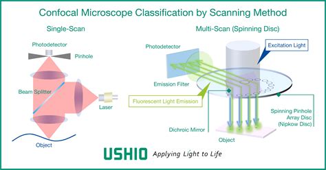
Measuring the thickness of objects which are labeled throughout is less accurate. Length and 2D area measurements are common image analysis problems and easily carried out with image analysis software. . both of which offer superior accuracy to confocal microscopes but can only measure surfaces. In the confocal microscope various types of .The sensors are ideally suited for high-precision distance and thickness measurements. The IFD2415 can also be used for multi-layer thickness measurements of up to 5 layers. The active exposure time control allows stable measurements on varying surfaces, even with dynamic processes of up to 25 kHz. Read moreFluorescence and confocal microscopes operating principle. Confocal microscopy, most frequently confocal laser scanning microscopy (CLSM) or laser scanning confocal microscopy (LSCM), is an optical imaging technique for increasing optical resolution and contrast of a micrograph by means of using a spatial pinhole to block out-of-focus light in image formation. [1] As a fast, high-accuracy and non-contact method, chromatic confocal microscopy is widely used in micro dimensional measurement. In this area, thickness measurement for transparent specimen is one of the typical applications. In conventional coaxial illumination mode, both the illumination and imaging axes are perpendicular to the test specimen. At the same .
Using this approach, the accuracy of confocal microscopic measurements was compared to tissue thickness measurements using ultrasound, a stan- dard method of measuring corneal thickness. 15 In a series of enucleated eyes from different species showing a range of corneal thickness from 300 to 700/~m, there was a significant correlation between . Mean corneal thickness measured by confocal microscopy was 516 ± 30 μm (±SD). This was less than the mean thickness measured by both ultrasonic pachymeters, 554 ± 28 μm by the DGH, and 555 ± 28 μm by the Sonogage (P < .001).Thickness measured by the Orbscan II pachymeter was 540 ± 35 μm (P < .001, compared with either confocal or .
Purpose: To quantify keratocyte density according to stromal region and subject age and to measure the thickness of the normal human cornea and its layers in vivo. Methods: Seventy normal corneas of 70 subjects were examined by confocal microscopy (contact lens wearers were excluded). Ages of subjects ranged from 12 to 80 years, with 10 subjects per decade.Save time on set-up as there is no need to change microscopes. Execute non-contact confocal imaging when evaluating surface roughness. LSM 900 is the ideal tool for a multi-user facility. Extend your upright light microscope, ZEISS Axio Imager.Z2m or your inverted light microscope ZEISS Axio Observer 7, with a confocal scanning module. Furthermore, the thickness measurement range always exceeds 1 μm to distinguish the peaks of the reflected spectrum. . Accurate and efficient height extraction in chromatic confocal microscopy using corrected fitting of the differential signal. Precis Eng, 56 (2019), pp. 447-454. Measuring the thickness of objects which are labeled throughout is less accurate. Length and 2D area measurements are common image analysis problems and easily carried out with image analysis software. . both .
In this paper, a new method for measuring the thickness of transparent specimens using chromatic confocal microscopy (CCM) is presented. The conventional CCM thickness measurement model relies on capturing the focal points on the upper and lower surfaces of a transparent specimen. This model has str . PDF | Accurate overlapping-peaks extraction plays a critical role in chromatic confocal thickness measurement of ultra-thin transparent film. However,. | Find, read and cite all the research you .
the setup like the spinning disk in the confocal microscopy.PSIRan be used for thickness measurement of solid and liquid transparent ˜lms in the range of micron to millimeter,here other methods .
These methods, however, require the lateral extent of the layer to be large, whereas we needed to measure the thickness of the layer in the small areas between pits (Fig. 2), so that an imaging system was necessary. The three-dimensional imaging capability of confocal microscopy offers an alternative approach to measurement of the layer thickness. This working principle is used for example in confocal microscopes [1,2], endoscopy [3], thickness measurement [4, 5], surface profiling [6] or tunable light sources [7]. To achieve high system .Mean corneal thickness measured by the Scheimpflug camera was 569 ± 52 μm (mean ± SD) and mean thickness measured by the confocal microscope was 570 ± 37 μm. These were not significantly different from each other ( P = 0.79, minimum detectable difference = 14 μm). Chromatic confocal microscopy is a widely used method to measure the thickness of transparent specimens. In conventional configurations, both the illumination and imaging axes are perpendicular to .
Scientific Reports - Thickness measurement of transparent liquid films with Paraxial Self-Reference Interferometry. . confocal microscopy can be used for thicknesses up to a few hundred microns . Chromatic confocal microscopy is a widely used method to measure the thickness of transparent specimens. In conventional configurations, both the illumination and imaging axes are perpendicular to . Confocal microscopy has been used to measure the wall thickness of microbubble cavities [31]; however, the image resolution limits the accuracy of the measured thickness to half a wavelength .
To measure corneal thickness by using a calibrated confocal microscope and to compare this measurement to thickness determined by ultrasonic and noncontact scanning slit pachymetry.
thickness of confocal scanning
quantitative confocal microscopy procedure
quantitative confocal microscopy guidelines

web20 de fev. de 2024 · 4.4K curtidas,124 comentários.Vídeo do TikTok de Rosy Aventura ® (@rosyaventura): "Parte 1 : #foryou #criança #kid #spiritbox #rosyaventura #sobrenatural #terror".Spooky, .
confocal microscopy thickness measurement|confocal fluorescent microscope metrics