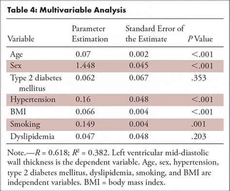
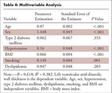
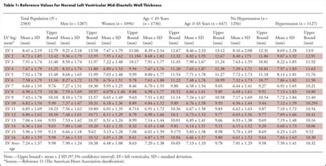
left ventricular wall thickness measurement|left ventricular hypertrophy with repolarization abnormality : traders Purpose. To generate normal reference values for left ventricular mid-diastolic wall thickness (LV-MDWT) measured by using CT angiography. Materials and Methods. LV-MDWT was measured in 2383 consecutive . WEB27 de ago. de 2019 · As informações e o palpite para Nice x Olympique de Marselha . Chega ao final nesta quarta-feira, 28 de agosto, a terceira rodada da versão 2019/2020 do Campeonato Francês. Com aproveitamento de 100%, o Nice recebe na Allianz Riviera, em Nice, o Olympique de Marselha, em confronto que tem pontapé inicial agendado para .
{plog:ftitle_list}
web30 de jan. de 2024 · Atlas had a forgettable first half of the season, with 15 points from 17 games to finish 17th in the Apertura, just two points above rock-bottom Necaxa. Pachuca vs Atlas Head-to-Head and Key Numbers
Purpose. To generate normal reference values for left ventricular mid-diastolic wall thickness (LV-MDWT) measured by using CT angiography. Materials and Methods. LV-MDWT was measured in 2383 consecutive .GLS should be measured in the 3 standard apical views (apical 4 chamber, 2 chamber and long axis) and the average GLS should be reported. Normal values depend on several factors .
After manually contouring the epicardial (green line in A–D) and endocardial (red line in A–D) border, myocardial thickness was automatically acquired in 100 measurements per left ventricular wall using the 2 .

Each echocardiogram includes an evaluation of the LV dimensions, wall thicknesses and function. Good measurements are essential and may have implications for therapy. The LV dimensions must be measured . LVM is the acronym for Left Ventricular Mass. LV mass (LVM) is a vital prognostic measurement we obtain with echocardiography to manage hypertension. RWT is the acronym for Relative Wall Thickness and is an .
Our LV calculator allows you to painlessly evaluate the left ventricular mass, left ventricular mass index (LVMI for the heart), and the relative wall thickness (RWT). Read on and discover all the details of our LV . In clinical routine, transthoracic echocardiography (TTE) is the standard first-line technique and is commonly used for follow-up. In this study we examined how CMR-derived .
normal left ventricular wall thickness
Echocardiography offers a reliable and reproducible method for measuring left ventricular wall thickness and mass. Finally, ultrasound may provide an accurate method for .

For the clinician, an increase in LV mass is the hallmark of LVH. 16 It can be estimated by measurements of LV dimension and wall thickness made with 2-D or M-mode echocardiograms, according to the formula of Devereux . To compare transthoracic echocardiography (TTE) and cardiac MRI measurements of left ventricular mass (LVM) and maximum wall thickness (MWT) in .
With physiologic remodeling, left ventricular wall thickness rarely exceeds 15 mm and left ventricular cavity sizes tend to be larger compared with the typical left ventricular cavity sizes in HCM. 6 Diastolic .
Encourage the holistic interpretation of measurements – no single number should define normality or pathology. . LVMi, left ventricular mass index; LVPWd, left ventricular posterior wall thickness in diastole. The BSE has chosen to publish an upper reference limit only for wall thickness measures with no separate partitions for mild .
normal left ventricle wall thickness
We measure the external diameter of the left ventricle of a heart. Enter your patient's interventricular septal end-diastole measurement (IVSd) It is also measured with echo. We evaluate the thickness of the wall that is .
Reference limits and values and partition values of left ventricular function Women Men Reference range Mildly abnormal . Normal values for Doppler-derived diastolic measurements Age group (y) Measurement 16-20 21-40 41-60 >60 IVRT (ms) . Relative wall thickness, cm 0.22–0.42 0.43–0.47 0.48–0.52 ≥0.53 0.24–0.42 0.43–0.46 . Background—We sought to compare maximal left ventricular (LV) wall thickness (WT) measurements as obtained by routine clinical practice between echocardiography and cardiac magnetic resonance (CMR) and document causes of discrepancy. Methods and Results—One-hundred and ninety-five patients with hypertrophic cardiomyopathy (median .
Measurement discrepancies of left ventricular mass and maximum wall thickness between modalities are clinically significant in patients with Fabry disease because use of one modality over the other could affect the diagnosis of left ventricular hypertrophy in 29% of patients, eligibility for disease-specific therapy in 26% of patients, and . Purpose To generate normal reference values for left ventricular mid-diastolic wall thickness (LV-MDWT) measured by using CT angiography. Materials and Methods LV-MDWT was measured in 2383 consecutive patients, without structural heart disease, undergoing prospective electrocardiographically (ECG) triggered mid-diastolic coronary CT angiography. .
Cardiovascular magnetic resonance (CMR) can accurately measure left ventricular (LV) mass, and several measures related to LV wall thickness exist. We hypothesized that prognosis can be used to . Left ventricular maximum wall thickness (MWT) is a key imaging biomarker in hypertrophic cardiomyopathy, guiding diagnosis, risk stratification, and clinical management. 1–4 For diagnosis, hypertrophic cardiomyopathy is clinically defined by an MWT of at least 15 mm in one or more left ventricular myocardial segments in the absence of abnormal loading .
Left ventricular (LV) wall thickness was determined by magnetic resonance (MR) in 15 patients (7 controls and 8 patients with coronary artery disease). End-diastolic (ed) and end-systolic (es) wall thickness were measured in a short axis view perpendicular to the LV long axis. Wall thickness measure .The Left Ventricle 3 1. Measurement of LV Size 3 1.1. Linear Measure-ments 3 1.2. Volumetric Measure-ments 3 1.3. Normal Reference Values for 2DE 6 . RV = Right ventricular RWT = Relative wall thickness STE = Speckle-tracking echocardiography TAPSE = Tricuspid annular plane systolic excursion TAVI = Transcatheter aorticLeft ventricular mass can be further estimated based on geometric assumptions of ventricular shape using the measured wall thickness and internal diameter. [7] Average thickness of the left ventricle, . CT and MRI-based measurement can be used to measure the left ventricle in three dimensions and calculate left ventricular mass directly.Background—We sought to compare maximal left ventricular (LV) wall thickness (WT) measurements as obtained by routine clinical practice between echocardiography and cardiac magnetic resonance (CMR) and document causes of . 3 Hindieh et al Left Ventricular Wall Thickness Discrepancy in HCM was 0.5 mm (95% confidence interval [CI], −6.9, 7. .
lv wall thickness normal values
Example: Measurement end - Diastolic wall thickness (red) + LV diameter (green) . Left ventricular dimensions. Women Men Reference range: Mildly abnormal: Moderately abnormal: . Relative wall thickness, cm 0.22–0.42 0.43–0.47 0.48–0.52 ≥0.53 0.24–0.42 0.43–0.46LVM is the acronym for Left Ventricular Mass. LV mass (LVM) is a vital prognostic measurement we obtain with echocardiography to manage hypertension. RWT is the acronym for Relative Wall Thickness and is an .

Normal (reference) values for echocardiography, for all measurements, according to AHA, ACC and ESC, with calculators, reviews and e-book. Shop e-books; Account; . Visual assessment of regional wall motion (left ventricle) .Figure 1: Images used for left ventricular (LV) mid-diastolic wall thickness and LV mass measurements. (a) Prospective electrocardiographically gated CT angiography study in mid-diastolic phase of the basal, mid, and apical (from left to right) short-axis (SAX) plane with LV wall thickness caliper measurements Background—To use cardiovascular magnetic resonance to investigate left ventricular wall thickness and the presence of asymmetrical hypertrophy in young army recruits before and after a period of intense exercise training. Methods and Results—Using cardiovascular magnetic resonance, the left ventricular wall thickness was measured in all 17 segments . Left ventricular hypertrophy (LVH) is a condition in which there is an increase in left ventricular mass, either due to an increase in wall thickness or due to left ventricular cavity enlargement, or both. Most commonly, the left ventricular wall thickening occurs in response to pressure overload, and chamber dilatation occurs in response to the volume .
Background—To use cardiovascular magnetic resonance to investigate left ventricular wall thickness and the presence of asymmetrical hypertrophy in young army recruits before and after a period of intense exercise training. Methods and Results—Using cardiovascular magnetic resonance, the left ventricular wall thickness was measured in all 17 segments . Background—We sought to compare maximal left ventricular (LV) wall thickness (WT) measurements as obtained by routine clinical practice between echocardiography and cardiac magnetic resonance (CMR) and document causes of discrepancy. Methods and Results—One-hundred and ninety-five patients with hypertrophic cardiomyopathy (median .Automate standard LV wall thickness measurements: Tag(s) #LV Functional Assessment: Panel. Cardiac. Define-AI ID. 18040013. Originator. . License. Creative Commons 4.0. Status: Published: Clinical Implementation. Value Proposition The measurement of left ventricular (LV) function is a well-established clinical parameter that has fundamental .In addition to the presence of LVH, the degree of ventricular thickness also has substantial prognostic value in many diseases. 10-12 Ventricular wall thickness is used to risk-stratify patients for risk of sudden cardiac death and help determine which patients should undergo defibrillator implantation. 10 Nevertheless, quantification of .
Rubber Density Meter services
Left ventricular maximal wall thickness (LVMWT) is integral in the diagnosis and risk stratification of hypertrophic cardiomyopathy (HCM). Echocardiography (TTE) is the primary modality by which LVMWT is assessed; however, there are limitations in obtaining accurate and reproducible LVMWT. . (IQR 2, 5); maximum LVMWT range for a single .Background: We sought to compare maximal left ventricular (LV) wall thickness (WT) measurements as obtained by routine clinical practice between echocardiography and cardiac magnetic resonance (CMR) and document causes of discrepancy.
Let’s now review 6 pitfalls to avoid when measuring the left ventricular wall and chambers. Avoid RV Trabeculations. . Incorrect: Correct: The trabeculation was measured as part of the IVS measurement causing the thickness of the septum to be over estimated: The gains, TGCs and focus were adjusted to help clearly define the endocardial .Hypertrophic cardiomyopathy (HCM) is a primary myocardial disease that primarily affects left ventricular (LV) myocardium and is characterized by mild to severe thickening (concentric hypertrophy) of the LV wall (septum and/or free wall) and papillary muscles. The thickening may be global or regional.
Rubber Impact Resiliency Tester services
Resultado da What’s new in GeForce Experience 3.26. What’s new in GeForce Experience 3.26. Support for Portal with RTX. GeForce Experience is updated to offer full feature support for Portal with RTX, a free DLC for all Portal owners.This includes Shadowplay to record your best moments, .
left ventricular wall thickness measurement|left ventricular hypertrophy with repolarization abnormality