standing twist test for mcl tear|mcl tear symptoms : manufacturing A medial collateral ligament (MCL) knee injury is a traumatic knee injury that typically occurs as a result of a sudden valgus force to the lateral aspect of the knee. Diagnosis can be suspected with increased valgus laxity . WEBpeituda novinha peituda gordinha peituda morena peituda goticas coroa peituda loira peituda negra peituda peituda gostosa branquinha peituda novinha peituda gostosa ruiva peituda. Goticas Peituda photos & videos. EroMe is the best place to share your erotic pics and porn videos. Every day, thousands of people use EroMe to enjoy free photos and .
{plog:ftitle_list}
Caesars Slots può contenere anche pubblicità. Per giocare p.
physical therapy for mcl tear
You may also stretch or tear your MCL if your knee is suddenly pushed to the side, or if it twists or bends out too far. Other movements that can cause an MCL tear include: . This article will explore the causes, symptoms, and treatment options for MCL injuries, highlighting what patients need to know for effective rehabilitation. We'll also touch on . Your healthcare provider will describe your MCL tear as one of the following three grades: Grade 1: A grade 1 MCL tear is a mild tear in which less than 10% of fibers in your .
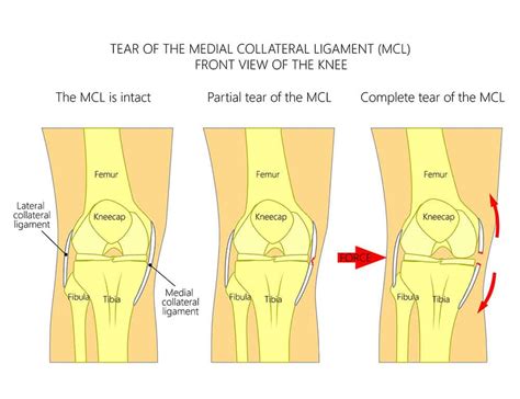
A medial collateral ligament (MCL) injury is a stretch, partial tear, or complete tear of the ligament on the inside of the knee. A valgus trauma or external tibia rotation are the causes of this injury.
A medial collateral ligament (MCL) knee injury is a traumatic knee injury that typically occurs as a result of a sudden valgus force to the lateral aspect of the knee. Diagnosis can be suspected with increased valgus laxity .
When the knee is hit on the outer side of the leg, or if the knee is twisted violently, the MCL can overstretch, causing a partial or complete tear. MCL injuries commonly occur in football . An MCL sprain is an excessive stretching or partial tear in the fibers of e the ligament. An MCL tear refers to a complete rupture of the ligament. A complete tear of the MCL can occur either where it attaches to the femur or to .Once the swelling and pain have lessened, your doctor will make the diagnosis. Your doctor may order a magnetic resonance imaging (MRI) scan. An MRI has an accuracy rate of nearly 90 .
Medial collateral ligament Injury of the knee (MCL Tear) are the most common ligament injuries of the knee and are frequently associated with ACL tears. They are cause by either a direct blow (more severe tear) or a non .
A doctor can usually diagnose an MCL injury based solely on the patient’s history and physical examination, with specific physical exam maneuvers that test the stability of the MCL, for example, keeping the femur stable while .Anatomy of the Meniscus [edit | edit source]. The meniscus is C-shaped cartilage that acts as a cushion between the proximal tibia and the distal femur to make up the knee joint. It has an average width of 10 mm to 12 mm, and the average thickness is 4 mm to 5 mm. The meniscus is made of fibro-elastic cartilage. It is an interlacing network of collagen, proteoglycan, .
Meniscus tears are the most common injury of the knee. Medial meniscus tears are generally seen more frequently than tears of the lateral meniscus, with a ratio of approximately 2:1. [2] Meniscal tears may occur in acute knee injuries in younger patients or as part of a degenerative process in older individuals. On physical exam, there is a mild effusion, medial joint line tenderness, and a positive medial McMurray test. Valgus stress reveals no pain or joint opening. Anterior and posterior drawer test is negative. . medial collateral ligament tear. meniscus tear. Associated conditions > 30% associated with anterior cruciate ligament injury . standing at 20 degrees of knee flexion on the affected limb, the patient twists with knee external and internal rotation with positive test being discomfort or clicking. McMurray's test flex the knee and place a hand on medial side of knee, . You will stand on one leg and twist side-to-side to identify any pain or other symptoms you feel during the movements. The Thessaly test is usually part of a preliminary exam when you visit your provider with knee pain or after an injury. You’ll probably also need at least one of a few imaging tests to confirm a torn meniscus or any other injuries in your knee.
This test is typically performed at both 30 and 0 degrees of knee flexion. When performed at 30 degrees, the MCL is more isolated from other medial joint structures, with a sensitivity of .86-.96 for MCL tears. This can be followed by performing the second version of the test, at 0 degrees of knee flexion, which allows for assessment of other . The medial collateral ligament (or MCL for short) connects the thigh bone (femur) to the shin bone (tibia) on the inside of your knee. It provides stability to your knee, preventing it from moving sideways. In particular from forces applied to the outside of the knee. The MCL itself has two parts to it:

mild mcl tear recovery time
Surgery for medial collateral ligament injury. Most people recover from an MCL injury without needing to have surgery. But sometimes, surgery is the best option to repair an injury to the medial collateral ligament. This is most likely if: more than one ligament or tissue in your knee is damaged; your knee remains unstable after physiotherapy Medial collateral ligament (MCL) injuries, like tears, can result in pain on the inside of the knee and instability. But don’t panic. While an MCL tear may sound scary, this injury is more often successfully treated with non-surgical options, like physical and exercise therapy, than other ligament injuries.The medial collateral ligament is injured more often than the lateral collateral ligament. Stretch and tear injuries to the collateral ligaments are usually caused by a blow to the outer side of the knee, such as when playing hockey or football. What are the symptoms of .Watch out for these MCL injury symptoms. MCL tears have similar symptoms to ACL tears, with some key exceptions. A blow to the inner knee can cause an MCL tear. Like the ACL, the injured person will feel pain, swelling, and tenderness. Unlike the ACL, there is no popping sound with the MCL. The pain will be specific to the inner knee with some .
Medial collateral ligament assessment (valgus stress test) The medial collateral ligament (MCL) assessment involves the application of a valgus force to assess the integrity of the MCL of the knee joint. The instructions below are for examining the right knee, use the opposite hands if assessing the left knee. 1.
An MCL tear is a painful but treatable condition, and most athletes return to their normal activities within a few weeks. Menu. Find a Doctor. Back. . Since MCL injury symptoms can be confused with symptoms of other medical conditions, your doctor may want to confirm an MCL tear with an X-ray, MRI or other test.What Are MCL Tears? An MCL tear is an injury to a ligament on the inside of the knee. MCL is an abbreviation for medial collateral ligament.The MCL is a strand of tough, fibrous tissue that connects the lower end of the thighbone or femur to the upper end of the shinbone or tibia.Its primary function is to stabilize the knee in standing and movement. To determine whether a tear is partial or complete, a doctor will perform several manual tests and order an MRI. Tests include: Lachman test: The physician will try to pull the shin bone away from the thigh bone. If the . Athletes and weekend warriors are the most susceptible to an MCL injury due to the nature of the physical contact they’re subjecting themselves to. Football, hockey, soccer, and other high impact athletic events which can cause an MCL knee injury. That said, an MCL sprain or tear can also happen with any general trip, fall, or even a slip. So .
The medial collateral ligament (MCL) is located on the inner aspect, or part, of your knee, outside the joint. Injury to the MCL is often called an MCL sprain or tear. MCL injuries are common in . The main function of the MCL is to resist a valgus force, in other words to prevent the tibia (shin bone) from moving too far outwards in relation to the knee joint. So when you are standing and your knee moves inwards, the MCL takes up the tension to prevent excessive movement.The medial collateral ligament is more vulnerable when the knee is bent, .
Valgus Stress Test for Medial Collateral Ligament Injuries. . As a stand-alone test, it had a sensitivity of 78% and specificity of 67% the pain was used as the outcome measure and a sensitivity of 91% and specificity of 49% when laxity was the outcome measure. So, the absence of both pain and laxity seems to be of at least moderate clinical . Rest the knee. Stop the activity that caused the injury. Limit movement to walking if the knee is painful. Use crutches to help relieve pain. Ice your knee to reduce pain and swelling. Do it for .The MCL ligament allows the knee joint to move but at the same time remain stable, preventing it from moving side to side. An injury to your MCL can range from a mild sprain or partial tear to a complete grade 3 rupture. A torn MCL can be painful, impair your ability to walk, and make it feel like you can’t hold your weight.

The medial collateral ligament's main function is to prevent the leg from extending too far inward, but it also helps keep the knee stable and allows it to rotate. Injuries to the medial collateral ligament most often happen when the knee is hit directly on its outer side. The medial collateral ligament usually responds well to nonsurgical treatment.
A “popping” sound when the injury occurs.This sound is usually a sign of a grade II or grade III tear. Immediate sharp pain from the inner section of the knee.; Immediate swelling at the inner knee. Swelling may increase and spread to the actual knee joint 1 or 2 days following injury.
The test is performed with the patient in standing with full weight bearing on the side to be tested. The foot should be flat on the floor. . Increased laxity compared to the unaffected side is considered a positive test for medial collateral ligament (MCL) injury. Varus stress test for Lateral Collateral Ligament.The medial collateral ligament (MCL) and lateral collateral ligament (LCL) are on the sides of the knee and prevent the joint from sliding sideways. . Acutely, the injury is a twist; the cartilage that is attached to and lays flat on the tibia is pinched between the femoral condyle and the tibial plateau. . Standing X-rays of the knees are .
We would like to show you a description here but the site won’t allow us.
standing twist test for mcl tear|mcl tear symptoms