tear test fluorescein stain and tonometery|ophthalmic test for tear stain : chain store Tear Break Up Time (TBUT) is used to evaluate the tear film quality. To perform this test, apply fluorescein stain to the eye, allow the animal to blink its eyelids, then examine with a cobalt blue light for areas of drying, which appear as dark spots. o Normal TBUT is 20 +/- 5 seconds o . ️
[email protected]
{plog:ftitle_list}
Resultado da Open Championship 2022: All the odds on three-time winner Tiger Woods. Join View market. Home. Golf. The Open. See the latest The Open Championship betting tips and previews from the expert .
Tear Break Up Time (TBUT) is used to evaluate the tear film quality. To perform this test, apply fluorescein stain to the eye, allow the animal to blink its eyelids, then examine with a cobalt blue light for areas of drying, which appear as dark spots. o Normal TBUT is 20 +/- 5 seconds o .Components of the minimum ophthalmic data base include: menace response, direct and consensual pupillary light reflex, palpebral reflex, Schirmer tear test, fluorescein stain, and . These tests include fluorescein staining, a Schirmer Tear Test, and tonometry. Keep reading to learn how these tests help your vet diagnose your pet’s eye problem. Fluorescein Staining; When might my vet use .Routine baseline tests like the Schirmer tear test (STT), fluorescein staining, and tonometry (intraocular pressure [IOP] measurement) are generally performed next. An STT should be .
What you can expect from tonometry tests can vary depending on the method. If you’re having applanation tonometry, your provider will add eye drops containing an anesthetic and a dye . Dr. Quintero suggests Fluress (fluorescein sodium and benoxinate hydrochloride ophthalmic solution, USP, 0.25%/0.4%, Akorn) contains too much fluorescein to be of value .
The proper order for the most common ocular tests are Schirmer Tear Test (STT), Fluorescein stain, and Intraocular pressures (IOP). 1) Schirmer Tear Test- The STT tests for dry eye or kerratoconjunctivitis sicca (KCS). This . The minimum ophthalmic database should include a complete ophthalmic exam, neurophthalmic exam, Schirmer tear test, tonometry, fluorescein stain, and additional diagnostics based on exam findings. Every .

Measurement of tear production is an important diagnostic test when deficiency of the lacrimal system is suspected. The tear-producing system is evaluated qualitatively by examination of . During GAT and Perkins tonometry, it is important to instill the correct amount of fluorescein in the eye, using either Fluress (fluorescein sodium and benoxinate hydrochloride, Akorn) or fluorescein strips with a .Applanation tonometry; Perforating injury - Seidel's test: If a perforation and active leak exist, the fluorescein is diluted by the aqueous and will appear as a yellow-green stream (of the diluted dye) within the dark orange (concentrated) dye. This is best appreciated while using the blue filter on the slit lamp. . A larger amount of stain .
In conclusion, ophthalmic exams play a crucial role in the diagnosis and management of various eye conditions in pets. Tests such as the Schirmer tear test, fluorescein test, tonometry, and ophthalmoscopy, as well as the . Fluorescein is a diagnostic contrast agent particularly used in various ophthalmic procedures, such as checking for any corneal or vessel abnormalities. The application of fluorescein also extends to bioimaging whole anatomic structures and even further to cellular components in immunohistological staining. This article outlines the indications, mechanism of . A fluorescein eye stain test can help your doctor detect corneal injuries, small foreign objects or particles in the eye, and abnormal tear production. . and abnormal tear production. The test .
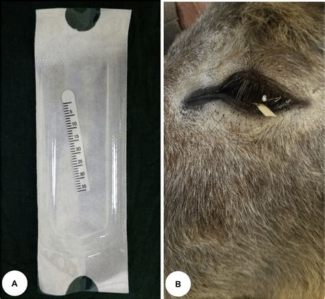
The tear film breakup time (TFBUT) test is a noninvasive assessment of precorneal tear film stability that is commonly performed when conjunctival hyperemia, chemosis, ocular discharge, or keratitis are noted. 4 In general, this test is performed by a veterinary ophthalmologist and requires fluorescein stain, a cobalt filter, and slit-lamp . Skill #18 Indications/reasons to perform Schirmer tear tests, fluorescein stains, and tonometry. A Schirmer tear test is important to perform in order to tell if the animal is producing enough tears in order the keep the eye healthy and also to detect dry eyes. This test can also help to determine any problems with the tear duct.

The GAT is mounted on the slit lamp to produce a magnified image, and when it touches the tear film and the cornea, a circle appears as to the observer. Staining the ocular surface with fluorescein and using a cobalt blue light in the slit lamp allow obtaining a better observation of the tear film(Fig.4). The final image looks like two .
Components of the minimum ophthalmic database include: menace response, direct and consensual pupillary light reflex, palpebral reflex, Schirmer tear test, fluorescein stain, and tonometry. Overview Ophthalmic emergencies are commonly seen by the small animal practitioner and can be said to include any ophthalmic condition that has rapidly .At this diameter, the resistance of the cornea to flattening is counterbalanced by the capillary attraction of the tear film meniscus for the tonometer head. The IOP (in mm Hg) equals the flattening force (in grams) multiplied by 10. Fluorescein dye is placed on the patient’s eye to highlight the tear film.Investigation of fluorescein stain–based tear film breakup time test .
schirmer tear test strips
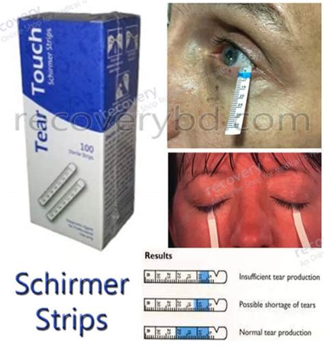
Measurement of tear production (using a strip of paper called the Schirmer tear test) Measurement of the pressures in the eye (tonometry ) Application of fluorescein, a green dye that checks for .
(See Schirmer Tear Test & Fluorescein Stain.) The Schirmer tear test (STT) measures basal tear production and reflex tear response and is often used to diagnose keratoconjunctivitis sicca (KCS). cl,ss="intro-deck"> 1sup> Basal tears are normal tears produced daily to lubricate and nourish the ocular surfaces. cl,ss="intro-deck"> 2sup>Fluorescein sodium stain is a hydrophilic dye used to evaluate tear film stability (tear film breakup time), integrity of the corneal epithelium (ulcers), corneal integrity (Seidel test), nasolacrimal duct patency (Jones test), and intraocular angiography. Fluorescence is intense under cobalt blue (450–500 nm) or ultraviolet light.Schirmer tear test (STT): Aids in diagnosis of conditions associated with decreased tear production, such as keratoconjunctivitis sicca (KCS), and should be performed before any medications are administered to the ocular surface .
Only the stroma absorbs stain • Use: Tear breakup time, Corneal or conjunctival ulcers, The Seidel test, Patency of nasolacrimal apparatus or Jones Test . The siedel test • Detects leakage of aqueous humor. Liberal application of fluorescein, gently press cornea, look for green dye indicating aqueous leakage . Jones test
Tear production Schirmer tear test Ophthalmic dyes Fluorescein stain uptake Intraocular pressure Tonometry KEY POINTS Ocular tests will help confirm the diagnoses in dogs and cats with ocular disease. The use of medication can influence values of the Schirmer tear test and tonometry. To limit variation of values and for better trending .Schirmer Tear Test. . Tonometry. After administering numbing eye drops, a digital tonometer is gently touched to the patient’s cornea to determine pressure inside the eye. High pressure could be indicative of glaucoma, while low pressure may be the result of inflammation in the eye. . The fluorescein stain test is also used to evaluate . The various methods used in applanation tonometry involve contact of the cornea with the instrument tip or with a column of air. Thus open globe wounds and traumas are contraindications. Goldmann and Perkins's applanation tonometry involves administering fluorescein and local topical anesthesia. Schirmer's Tear Test must come first before any topical anesthetic or stain and preferably, hours after any topical ointment. . The fluorescein stain is available in strips or multidose vials. In our hospitals where the bottles are used so frequently we go through almost one a day, the chance of bacterial or fungal contamination of the .
This is a test that uses orange dye (fluorescein) and a blue light to detect foreign bodies in the eye. This test can also detect damage to the cornea. The cornea is the outer surface of the eye.Fluorescein Stain 2. Schirmer Tear test 3. Tonometry. What are opthalmoscopes used for? . What are normal values for a tonometry test in a dog? a cat? 15-20 mmHg dog 20-30 mmHg cat. What instrument is used to check the ears of a patient? Otoscope. 1. Handle 2. Connecting Piece 3. Otoscope head
Schirmer tear test: A test that measures the production of tears that protect the eye from debris; Fluorescein stain: A test that identifies any defects in the eye surface; Tonometry: Identifies the pressure within the eye to rule out glaucoma; Depending on the diagnosis, treatment plans include: Lubricating eye drops; Topical antibiotics
What you can expect from tonometry tests can vary depending on the method. If you’re having applanation tonometry, your provider will add eye drops containing an anesthetic and a dye called fluorescein to your eyes. But non-contact tonometry and most other methods don’t need either of these to work. Some of the most common methods for the .A fluorescein stain test is the most common eye test performed and may be the only test needed if the ulcer is acute and very superficial. If the ulcer is chronic or is very deep, samples may be taken for culture and cell study prior to applying the stain or other medication. . The tear ducts drain tears away from the eyes into the back of .
schirmer tear test pdf
KCS-schirmer tear test Corneal ulceration -fluorescein stain glaucoma -tonometry Uveitis -miosis, aqueous flare Immune mediated -diagnosis of exclusion. 1 / 7. 1 / 7. . KCS-schirmer tear test Corneal ulceration -fluorescein stain glaucoma -tonometry Uveitis -miosis, aqueous flare Immune mediated -diagnosis of exclusion. Uvea consists of: iris .
Tonometry is a standard procedure employed by ophthalmologists to measure intraocular pressure (IOP) using a calibrated instrument. Devices used to measure IOP are based on the assumption that the eye is a closed globe with uniform pressure distributed throughout the anterior chamber and vitreous cavity. The normal range of intraocular pressure is 10 to 21 .

web4 dias atrás · Sinopse o livro (resumo) «Um casamento arranjado» Zana Kheiron. Carolina Navarro será obrigada por seu pai a se casar com um homem desfigurado, a fim de salvar a família da ruína. Máximo Castillo tinha tudo o que qualquer um poderia querer, até que um acidente de avião destruiu seu corpo, sua alma, seu relacionamento, tornando-o .
tear test fluorescein stain and tonometery|ophthalmic test for tear stain